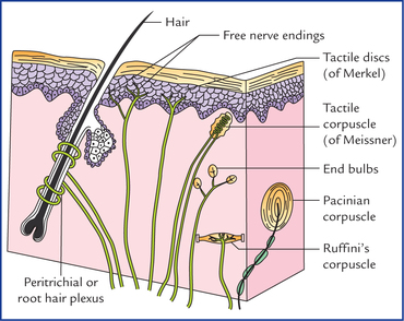
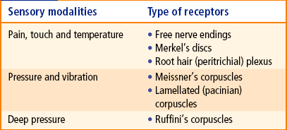
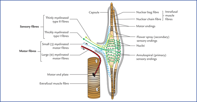
4 Whenever a clinician performs a neurological examination in a patient, he/she tests for normal function of the sensory input and motor output so that he/she may find out any sensory or motor deficit if it is there. The knowledge of structure and function of sensory and motor nerve endings responsible for sensory input and motor output is essential while performing these tests. An individual receives information from outside and from within the body by special sensory nerve endings called receptors. The receptor receives stimulus and converts it into a nerve impulse. The receptors thus act as transducers,* converting mechanical and other stimuli into electrical impulses. Thus, receptors are sensory nerve endings specialized for reception of stimuli and transmitting them in the form of nerve impulses. The receptors can be classified broadly into: • Exteroceptors: They provide information of touch, pain, temperature and pressure. These are superficially located, such as in skin, and are also called cutaneous receptors. • Proprioceptors: They provide information about state of contraction of muscles and of joint movement and position. • Interoceptors: They provide information from viscera and blood vessels. • Mechanoreceptors: They are stimulated by mechanical deformation. • Chemoreceptors: They are stimulated by chemical influences. • Thermoreceptors: They respond to alternation in temperature, e.g. cold and heat. • Nocireceptors: These respond to any stimuli that bring about damage to the tissue. Damage to tissue is perceived as pain, discomfort or irritation. • Photoreceptors: They are stimulated by light, e.g. rods and cones of retina. • Osmoreceptors: They respond to changes in the osmotic pressure. Structurally the receptors are classified into two types: non-encapsulated and encapsulated. • Free nerve endings (Fig. 4.1): They are widely distributed in the body tissues such as skin, cornea, periosteum, dental pulp, etc. The afferent fibres from free nerve endings are either myelinated or non-myelinated. The terminal endings are devoid of a myelin sheath and there are no Schwann cells covering their tips. Most of these endings carry pain sensations (pain fibres), but they are also sensitive to temperature, touch, pressure and tickle sensations. • Peritrichial or root hair plexus (Fig. 4.1): It is a network of dendritic branches that surrounds the outer root sheath of hair follicles and is stimulated by light touch causing movement of hair. • Tactile discs (of Merkel): They are expanded disc-like nerve endings in the germinative epidermal layer of hairless skin (Fig. 4.1). They make close contact with Merkel cells, which are specialized epithelial cells in the deeper part of the epidermis. The tactile discs are slowly adapting touch receptors that transmit information about the degree of pressure exerted on skin, e.g. when one is holding a pen. Here the sensory nerve endings are enclosed in a capsule derived from surrounding non-neural cells. • Tactile corpuscles (of Meissner): They are ovoid in shape and found in the dermal papillae of the skin in those areas where tactile sensitivities are extremely well developed, viz. eyelids, lips, fingertips, nipples and external genitalia. The corpuscle consists of a capsule and a central core. The central core contains epithelioid cells (modified Schwann cells) and is supplied by several myelinated nerve fibres. The capsule is continuous with the perineu-rium of nerves supplying the corpuscle. They function as low threshold rapidly adapting mechanoreceptors and are responsible for close spatial ‘two point’ discrimination (i.e. they enable an individual to distinguish between two pointed structures when they are placed close to each other on the skin). • Pacinian corpuscles (Fig. 4.1): They are the largest and most numerous encapsulated receptors. Each corpuscle is ovoid in shape, measuring up to 2 mm in length and consists of an outer laminated capsule of flat cells which are arranged in concentric layers like an onion peel and a central core. A myelinated nerve fibre loses its sheath to enter its central core. Pacinian corpuscles are rapidly adapting mechanoreceptors that are particularly sensitive to firm pressure (pushing) and vibration. These are scattered throughout the integument of the body notably in the subcutaneous tissue of palm, sole, fingers and breasts. • End bulbs of various types: They consist of multiple branched nerve terminals, which are encapsulated (Fig. 4.1). – Bulbous corpuscles (of Krause) are spherical and found mainly at the mucocutaneous junctions. – Genital corpuscles (or Golgi-Mazzoni) are slightly different from bulbous corpuscles (of Krause) and occur in the genital skin. • Ruffini’s corpuscles: They are spindle-shaped structures located into the dermis of hairy skin. Each corpuscle consists of several non-myelinated nerve endings of a large myelinated axon within a bundle of collagen fibres and surrounded by a cellular capsule. They are slowly adapting mechanoreceptors which respond when skin is stretched causing stresses in dermal collagen. Hence, these are stretch receptors like Golgi tendon organs. The skin contains both non-capsulated and encapsulated receptors such as free nerve endings, Merkel’s discs, root hair plexus, Meissner’s, Pacinian and Ruffini’s corpuscles. Various sensory modalities detected by different receptors in skin are listed in Table 4.1. The muscle receptors comprise neuromuscular spindles (Fig. 4.2). These are spindle-shaped sensory end organs found in the skeletal muscles and are most numerous towards the tendinous attachment of the muscle. They provide sensory information to the CNS to control the motor activity and tone of the muscle. Each spindle consists of a bundle of small specialized skeletal muscle fibres (3–10), and is surrounded by a fusiform capsule of connective tissue. The specialized muscle fibres within the capsule are called intrafusal fibres and ordinary muscle fibres situated outside the spindle are referred to as extrafusal fibres. The slender intrafusal fibres are striated only at the ends; therefore, only the ends of these fibres can contract.
Receptors and Effectors
Receptors
Classification of Receptors
Functional types
On the basis of the kind of information they provide
On the basis of the manner in which they are stimulated
Anatomical types
Non-encapsulated receptors
Encapsulated receptors
Cutaneous Receptors (Fig. 4.1)
Muscle Receptors
Neuromuscular spindles
![]()
Stay updated, free articles. Join our Telegram channel

Full access? Get Clinical Tree


Receptors and effectors