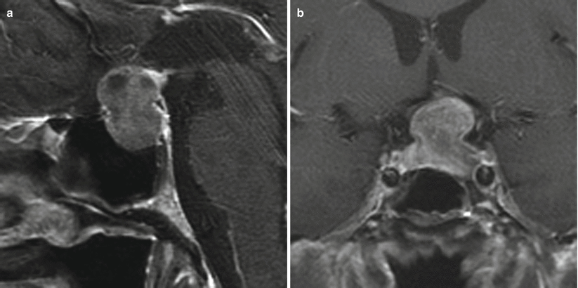
Fig. 14.1
Silent corticotroph (ACTH) adenoma. (a, b) Sagittal T1-weighted post-gadolinium image. (c) Coronal T1-weighted post-gadolinium image. There is a large, heterogeneously enhancing sellar mass with suprasellar extension and encasement of bilateral cavernous internal carotid arteries suggestive of invasion. There is erosion of the clivus. The normal pituitary gland appears along the superior margin of the mass

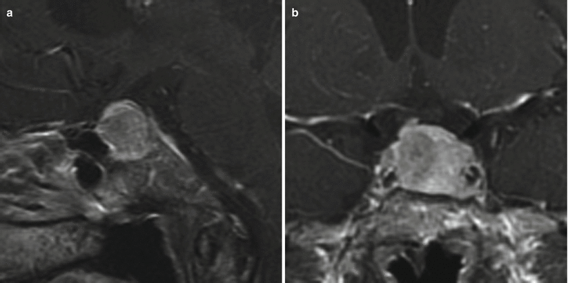
Fig. 14.2
Silent corticotroph (ACTH) adenoma. (a) Sagittal T1-weighted post-gadolinium image. (b) Coronal T1-weighted post-gadolinium image. A heterogeneously enhancing sellar/suprasellar mass contacts the left cavernous internal carotid artery without encasement. The optic chiasm is elevated by the mass

Fig. 14.3
Silent corticotroph (ACTH) adenoma. (a) Sagittal T1-weighted post-gadolinium image. (b) Coronal T1-weighted post-gadolinium image. A heterogeneously enhancing sellar mass contacts bilateral cavernous internal carotid arteries with possible encasement on the left. The normal pituitary gland appears along the superior margin of the mass, with the stalk deviated to the right

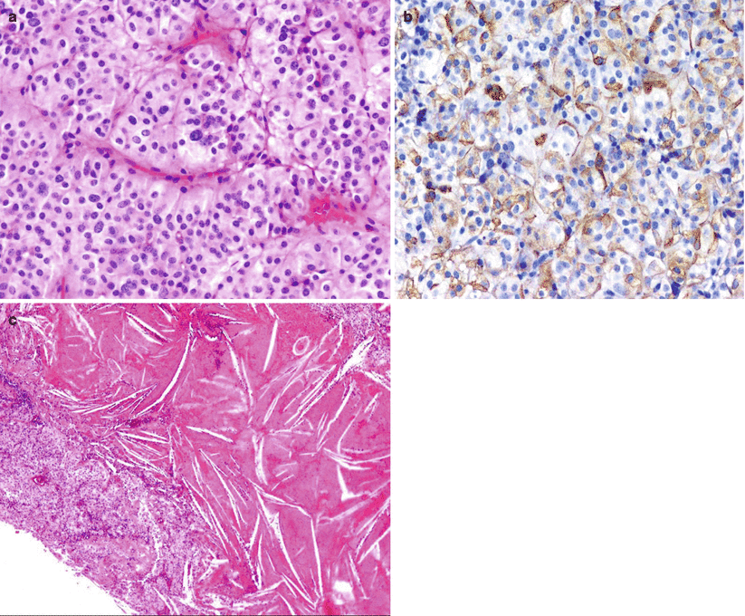
Fig. 14.4
Silent corticotroph (ACTH) adenoma that presented clinically as a nonfunctioning adenoma. (a) A chromophobic adenoma exhibiting a diffuse growth pattern. (b) Despite lack of clinical symptoms, this adenoma shows immunopositivity for ACTH. (c) The same adenoma displays a large area of previous hemorrhage with accumulation of cholesterol clefts. Silent corticotroph adenomas show a high tendency for hemorrhage and/or apoplexy, which may be the presenting symptom in about one third of patients
14.2.2 Histopathology
Stay updated, free articles. Join our Telegram channel

Full access? Get Clinical Tree


