Fig. 45.1
Sinus malignancy: melanoma. (a) Sagittal CT image with contrast enhancement. (b) Sagittal T1-weighted gadolinium-enhanced MR image. (c) Coronal T1-weighted gadolinium-enhanced image. A heterogeneously enhancing mass is centered in the sphenoid sinus, eroding the upper clivus. The pituitary gland is immediately along the superior/posterior margin of the mass
45.3 Histopathology
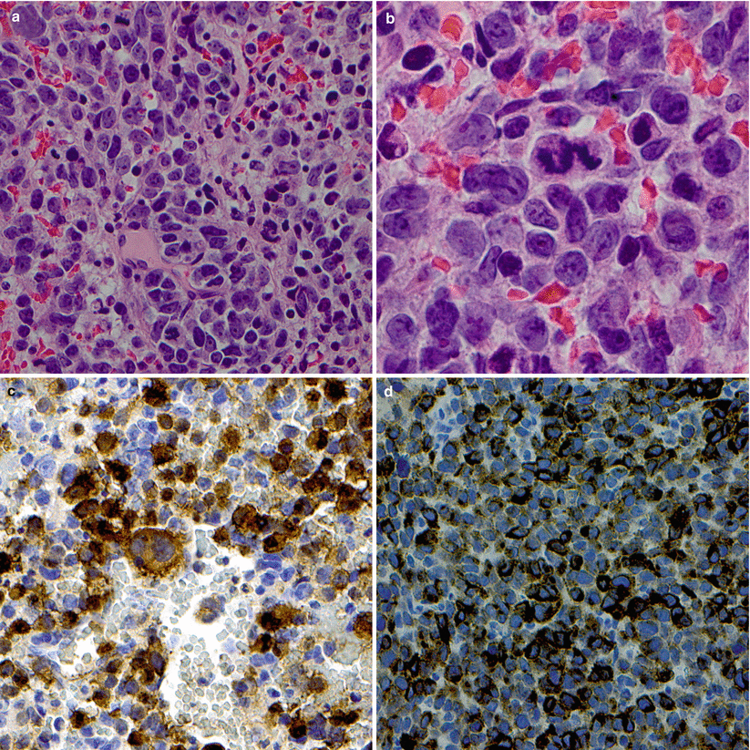
Melanoma is characterized as a malignant melanocytic tumor with immunopositivity for S100 protein and HMB-45 (Fig. 45.2).
Common histopathological features include necrosis, pigmentation, vascular invasion (68 %), and a lymphocytic response (63 %) [15].
Tumor pigmentation and papillary pseudoarchitecture are associated with a worse outcome [16].

Fig. 45.2
Sinus malignancy: melanoma. H&E staining demonstrates epithelioid-shaped cells with numerous mitoses, prominent “cherry-red” nucleoli, and weakly eosinophilic and slightly blue-gray cytoplasm (a). This lesion had associated hemorrhage and microhemorrhage (×40 objective). A high-power view (b) demonstrates the same features (×100). Malignant cells were stained positively by anti-S100 antibodies (c) and by anti-MelanA/MART1 antibodies (×20) (d). Binucleate forms may also be seen in melanoma (c)
45.4 Clinical and Surgical Management
A standard TNM classification system is used to stage patients and has been shown to correlate with outcomes [17].
Maximal safe tumor debulking is recommended, followed by adjunctive chemotherapy, radiation, or both. When possible, obtaining tumor-free margins is recommended.
Chemotherapy has been shown to offer a greater survival benefit than radiation [18].
Current options for chemotherapy include dacarbazine, temozolomide, paclitaxel, and others. Chemotherapy may be administered with or without interferon-alpha.
Craniofacial approaches have traditionally been used to achieve maximal safe tumor resection [4].
Endoscopic approaches have become the mainstay of surgical treatment for many patients with skull base melanoma [19, 20].
Recurrence and the development of distant metastases are common.
References
1.
2.
3.
Clifton N, Harrison L, Bradley PJ, Jones NS. Malignant melanoma of nasal cavity and paranasal sinuses: report of 24 patients and literature review. J Laryngol Otol. 2011;125:479–85.CrossRefPubMed
Stay updated, free articles. Join our Telegram channel

Full access? Get Clinical Tree


