Fig. 47.1
Sinonasal undifferentiated carcinoma (SNUC). (a) Sagittal T1-weighted gadolinium-enhanced MR image. (b) Coronal T1-weighted gadolinium-enhanced image. A large, heterogeneously enhancing mass involves the nasal cavity and ethmoid and sphenoid sinuses, invades the left orbit, and extends through the cribriform plate and left orbital roof to the left anterior cranial fossa, with displacement of the left frontal lobe
47.3 Histopathology
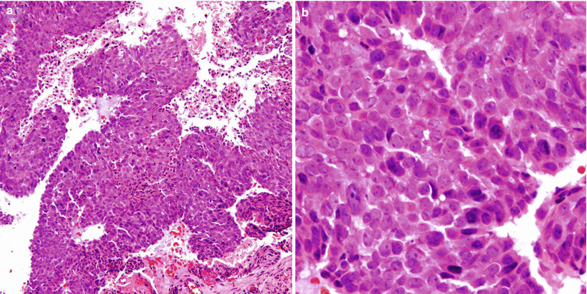
SNUC is a highly aggressive carcinoma characterized by medium-sized cells with small to moderate amounts of eosinophilic cytoplasm, frequent mitoses, necrosis, and prominent vasculature (Fig. 47.2) [2].
Positive immunoreactivity for pancytokeratins, simple keratin, EMA, and neuron-specific enolase is common [2].

Fig. 47.2
SNUC. (a) SNUCs are composed of nests and lobules of undifferentiated cells with no squamous or glandular differentiation. Necrosis may be prominent. (b) The cells are medium to large in size, with a large nucleus and prominent nucleoli
47.4 Clinical and Surgical Management
Both the Kadish staging system and Hyams grading system can be adapted to predict outcome in SNUC [7, 8].
Multimodal treatment consisting of maximal safe tumor resection, chemotherapy, and radiation is recommended and has been shown to extend patient survival [9].
Intensity-modulated radiation therapy (IMRT) is the recommended treatment modality for radiation [10].


