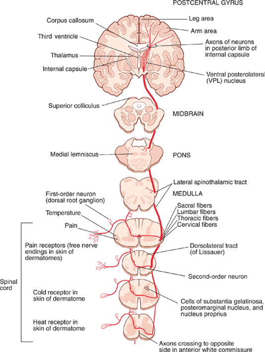
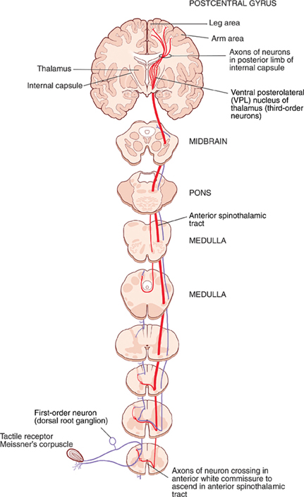
14 Somatosensory System Testing the somatosensory system constitutes one of the most difficult but most important aspects of the neurologic examination. A logical approach to this examination requires an understanding of the basic modalities of sensation, the major fiber tracts that they follow, and the primary fiber types in which they are carried. This chapter describes two major components of the somatosensory systems: (1) the spinothalamic system, which is responsible for pain, temperature, light touch, and pressure sensation; and (2) the dorsal column—medial lemniscus system, which is responsible for position sense, vibratory sense, and discriminative touch. Sensory pathways for the face and cortical processing of sensory information are also described. The spinothalamic system may be divided into two sensory pathways: the lateral spinothalamic tract, which mediates pain and temperature sensation, and the anterior spinothalamic tract, which mediates light touch and pressure sensation. See Fig. 14.1. Sensory receptors that mediate pain and temperature sensation are known as nocireceptors and thermorecep-tors, respectively. Nociceptors (pain receptors) and ther-moreceptors (temperature receptors) are free nerve endings of axons in the A-delta and C fiber classes. A-delta fibers are slightly myelinated compared with C fibers, which are unmyelinated. The abrupt, sharp, and well-localized sensation of so-called fast pain, carried by A-delta fibers, is commonly followed by the noxious and burning sensation of slow pain, carried by C fibers. Pain and temperature sensation are carried in spinal pathways. The cell bodies of pain- and temperature-related fibers (A-delta and C fibers) are located in the dorsal root ganglia. These first-order neurons send central processes through the lateral portions of the dorsal roots to enter the dorsolateral tract (of Lissauer) of the spinal cord. In the dorsolateral tract these neurons ascend or descend one to three spinal segments from their level of entry and then synapse in the substantia gelatinosa (lamina II of the dorsal gray horn), the posteromarginal nucleus (lamina I), and/or the nucleus proprius (laminae III and IV). After synapsing in the dorsal horn, second-order neurons send axons to the contralateral lateral spinothalamic tract, often crossing through the anterior white commissure. Because the tract is formed by crossed fibers that are added medially as the spinal cord is ascended, the central fibers of cells (whose peripheral fibers supply the legs) are located lateral to those that supply the arms. Therefore, the spinothalamic tract is somatotypically arranged, with the upper extremities represented medially and the lower extremities and sacrum laterally. During its passage through the brainstem, the lateral spinothalamic tract is known as the spinal lemniscus. The spinal lemniscus is located lateral to the medial lemniscus and contains fibers of the spinotectal tract destined for the superior colliculus. The spinal lemniscus also gives off collateral branches to the reticular formation at several levels of the brainstem. Fibers of the second-order neurons of the lateral spi-nothalamic tract terminate in the ventral posterolateral (VPL) nucleus of the thalamus. The perception of pain and temperature is initiated in the VPL nucleus and later modified in the cerebral cortex. Somatotopic order is maintained in the thalamus. The cell bodies of third-order neurons are located in the VPL nucleus of the thalamus. These cell bodies project axons through the dorsal limb of the internal capsule and the corona radiata to reach the primary somatosensory cortex I in the postcentral gyrus (Brodmann’s areas 3, 2, and 1) and somatosensory area II in the anterior aspect of the parietal speculum. Sensory information is distributed from these areas to other regions of the cortex, including motor areas and the parietal association areas. Fig. 14.1 Lateral spinothalamic tract. See Fig. 14.2. Light touch and pressure sensation are mediated by specialized receptors in the skin. Among these are free nerve endings, such as Merkel’s cells, and encapsulated endings, such as Meissner’s corpuscles. The anterior spinothalamic tract runs ventromedially in relation to the lateral spinothalamic tract. First-order neurons located in the dorsal root ganglia send central processes though the dorsal roots that enter the dorso-lateral tract of the spinal cord, ascend or descend one to three segments, and synapse on dorsal horn cells. These dorsal horn cells, constituting the second-order neurons, project axons that traverse the anterior white commissure to contribute to the contralateral anterior spinotha-lamic tract, which maintains the same somatotopic order as the lateral spinothalamic tract. After ascending the spinal cord and traversing the brainstem (as part of the spinal lemniscus), these fibers terminate on third-order neurons in the VPL nucleus of the thalamus. Third-order neurons in the VPL thalamic nucleus project axons through the dorsal limb of the internal capsule and the corona radiata to reach the postcentral gyrus of the cerebral cortex.
Spinothalamic System
Lateral Spinothalamic Tract
Receptors
Spinal Pathways
Thalamocortical Projections

Anterior Spinothalamic Tract
Receptors
Spinal Pathways
Thalamocortical Projections

Stay updated, free articles. Join our Telegram channel

Full access? Get Clinical Tree


