Chapter 145 Spinal Infections
Vertebral Osteomyelitis and Spinal Epidural Abscess
Epidemiology of Spinal Infection
During recent decades, there has been a steady rise in the incidence of VO and SEA, traditionally felt to be relatively infrequent infections of the vertebral column. Although older series reported rates of VO and SEA of 0.2 to 2 per 10,000 hospitalizations, researchers in more recent series in both Europe and the United States have reported a 10- to 15-fold increase in the incidence of VO and SEA at large tertiary care centers.1–6
Additionally, though older series reported a biphasic distribution in the age-related incidence of VO and SEA, with incidence peaking in the first and fifth to sixth decades of life, newer series have shown a rising incidence of these infections in adult patients with impaired host defenses, frequent bacteremia, or both.7–10 In particular, in recent population-based reports, patients with AIDS and intravenous drug users have been shown to have exceedingly high relative rates of VO or SEA.11–13 Patients with diabetes, liver cirrhosis, renal disease on dialysis, and patients on chronic immunosuppression have all shown a proclivity toward the development of spinal infections.14 The widespread use of MRI technology, coupled with the greater concentration of these patients at tertiary care centers, may account for at least a portion of the greater relative incidence of these spinal infections documented in recent years.
Risk Factors for Vertebral Osteomyelitis and Spinal Epidural Abscess
Several risk factors have been associated with osteomyelitis and SEAs. Many of these risk factors are associated with an immunocompromised state and appear to lead to the development of the infection. Diabetes, for example, lowers the immune response and is associated with spinal infections. Studies have shown diabetes to be associated with SEAs in 18% to 54% of cases3 and 31% of VO cases.15 IV drug abuse has also been associated with the development of spinal infections. This may be secondary to the sharing of unsterile needles, leading to systemic infections and endocarditis, which then predispose individuals to spinal infection. There also seems to be an association between alcoholism and spinal infections. This association may have multifactorial causes, including diet and hygiene and increased predisposition to spinal trauma.16 Other associated risk factors that decrease immune system function include malignancy and long-term steroid use.
Since one of the major mechanisms of the development of VO and SEA include hematogenous dissemination of an infection from a remote source, infections and bacteria originating from other parts of the body serve as risk factors for spinal infections. Therefore, spinal infections have been associated with other infections, including bacterial endocarditis, urinary tract infections, skin abscesses, pneumonia, dental infections, and sepsis. In several studies, skin and soft-tissue infections were found to be the most common cause of SEAs via hematogenous spread.11,14 In VO, however, the most common source of infection seems to be from the urinary tract.17 Hematogenous seeding of spinal epidural infections account for approximately 50% of cases.18,19
In addition to hematogenous dissemination, SEAs may develop by direct extension from an area of VO. In such cases, the abscess tends to be located in the ventral epidural space. Several studies have shown this association of SEA with VO, especially in ventrally located abscesses. Infections extending to the spinal epidural space can also directly spread from a psoas muscle abscess. This route of spread is responsible for approximately 10% to 30% of cases.19
Spinal infections may also develop from direct seeding of bacteria via an invasive procedure. Open spinal surgery, including laminectomies, discectomies, and operations involving spinal instrumentation, as well as less invasive procedures, such as lumbar punctures and epidural injections, have been shown to be risk factors for the development of spinal infections. Interestingly, the rate of infection due to less invasive procedures seems to be larger than that due to open spinal surgeries. Researchers in recent studies have found the risk of developing a SEA after epidural catheterization to be from 0.04% to 0.07%.19 Moreover, leaving catheters and drains in place at the time of surgery seems to increase the risk of acquiring a spinal infection.18,19 Trauma to the spine has also been associated with the development of SEA and VO. Trauma may predispose patients to spinal infections because of disruption of anatomic barriers.18
Clinical Presentation
The classic triad of symptoms of SEA is fever, back pain, and neurologic dysfunction. Investigators in some studies, however, have reported that this triad is present in only 10% to 15% of patients at the time of initial presentation.18,19 Important features of the clinical presentation of SEA are that the symptoms progress through several stages and that this progression can occur rapidly. A classic paper by Heusner1 described the stages of clinical SEA presentation:
The course of time through which these stages progress is highly variable. A patient may present with symptoms that have been ongoing from days to weeks.19 An abscess caused by hematogenous spreading tends to have a clinical presentation that progresses more rapidly through these stages as compared to an abscess formed secondarily to local extension. Once neurologic dysfunction is present, however, the average time to complete paralysis is 24 hours and often can occur in even less time. Thus, the onset of neurologic dysfunction should prompt the need for emergent surgical decompression to prevent progression to paralysis.
Localized pain and tenderness over the involved area of the spine is found very commonly as the initial presentation of SEA (71% in one large meta-analysis).11 Oftentimes pain is described as sharp and stabbing19 This pain reflects the level of the spine involved. The pain may occur in a dermatomal distribution in the lower extremities, for example, or a patient may experience pain in the chest wall or abdomen which may mimic other conditions such as pancreatitis.19 Neurologic dysfunction secondary to a SEA may include motor weakness, including sphincter dysfunction and sensory loss. Motor deficits were shown to be present in 54% of cases in one study.20 A large meta-analysis showed paralysis affects approximately 34% of patients with SEA.11
Fever is also a common manifestation of SEA, with an incidence between 66% and 75%.16 Symptoms and signs of meningitis may also be present, including headaches and nuchal rigidity. This may represent a parameningeal reaction. In patients with a chronic infection course, symptoms such as unintended weight loss and malaise can occur. Sometimes the clinical presentation of SEA may be dominated by that of sepsis, making the detection of a SEA more obscure. In children, the symptoms are more likely to be nonspecific, and the child may simply complain of not feeling well.18,21
The clinical manifestations of VO are similar to those of SEA in that localized pain exacerbated by motion and tenderness almost always occurs and is very frequently the initial symptom. Often-times the pain may be associated with paraspinal muscle spasm. In one review, the authors reported a 92% incidence of back pain in VO.17 Radicular pain in VO has been reported with incidences of up to 25-33%.15 Neurologic deficits can occur with VO and are due to either associated SEA or spinal instability. Investigators in one study reported neurologic impairment in 28% of patients with VO.22 Neurologic involvement seems to be more common in cases of VO affecting the cervical or thoracic spine as compared to the lumbar spine.23 Fever can also be present in VO; however, this presentation is not as consistent a finding as back pain and tenderness.
Laboratory Findings
Laboratory findings associated with VO and SEA reflect the underlying inflammatory nature of the disease and are not specific. The erythrocyte sedimentation rate (ESR) is the most consistently elevated laboratory value in spinal infection, with the ESR typically being greater than 40. In one meta-analysis, the average ESR was 77.2,8 ESR has been shown to be greater than 20 in 95% of cases in some studies.3,19 The ESR is also helpful in monitoring the disease process during therapy, with declining values of ESR to be expected with appropriate therapy. The C-reactive protein (CRP), an acute phase reactant, has also been found to be elevated in patients with spinal infections.2,8 Obtaining CRP levels may be useful because CRP levels have been shown to rise more quickly during inflammation and to return to normal more quickly than ESR levels.24 In addition, one study showed CRP values obtained over 2 weeks after surgery for SEAs can provide prognostic information regarding outcomes.20
The peripheral white blood cell count may be elevated in patients with spinal infections, but many times may be normal. An elevation of the peripheral white blood cell count may be more indicative of a systemic infectious process. The average peripheral white blood cell count in one large meta-analysis of SEAs was 15,700/μL.11
In patients with SEA, a cerebrospinal fluid (CSF) sample may show an elevated white blood cell count and protein level, which are indicative of a parameningeal process. In addition, the CSF values may be reflective of concomitant encephalitis or meningitis, which may present along with a spinal infection in a significant proportion of cases.2 However, a lumbar puncture is often inadvisable in the setting of a SEA because of the risk of seeding bacteria into the thecal sac, especially in abscesses in the lumbar region that are located in the dorsal epidural space.
Blood cultures are often positive in the setting of a spinal infection. The organism grown on blood culture may be the same as the causative organism in a SEA or VO, especially if the spinal infection was caused by a hematogenous seeding. Blood cultures have been reported to be positive in approximately 50% of cases in some studies.15 Positive blood cultures can be useful for guiding antibiotic therapy in the setting in which direct cultures from the spinal infection cannot be obtained. Because blood cultures may become negative if obtained after the administration of antibiotics, it is important to obtain blood cultures prior to the initiation of antibiotics if possible.
Radiographic Studies
Narrowing of the intervertebral disc space can be seen in plain films as early as 2 weeks from the onset of symptoms but usually appear at week 6 or 8.15,26–28 As the infection progresses, there is an increase in vertebral body destruction followed, in some cases, by subsequent spinal deformity. Lytic lesions, demineralization, and scalloping of the end plates may occur.25 As the infection resolves and healing takes place, new bone formation and subsequent fusion can occur, but this may not be evident for as long as 1 year.
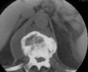
For the above-described reasons, computed tomography (CT) has become an important diagnostic tool in the diagnosis of spinal infections, especially for VO. Among its benefits, CT provides excellent detail of the bony anatomy, and it clearly demonstrates the lytic lesions as well as gas within the disc space or soft tissues (Fig. 145-1). Although identification of SEA has been reported using CT alone, it is generally thought that plain CT is not particularly sensitive for visualizing SEA.29 Indirect findings such as intraspinal gas have been described in the presence of SEA.25
CT is the most valuable tool when performing image-guided percutaneous biopsies of the vertebral body and paravertebral tissues to obtain culture material, and CT is also effective in performing aspiration of paravertebral abscesses.30 Image-guided CT biopsy has been shown to be safe and effective in obtaining diagnostic material at all levels of the spine; however, this technique yields a lower diagnostic rate than the previously reported biopsy of neoplastic vertebral lesions, especially if performed in patients who have undergone previous antibiotic treatment.31 Therefore, it is imperative to perform the biopsy prior to administration of antibiotics to maximize the yield of a positive culture.
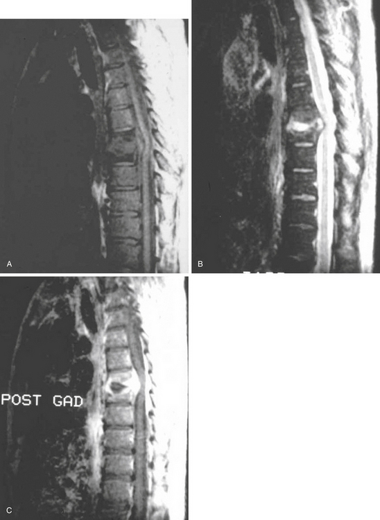
MRI has lately become the single most important imaging modality in the evaluation of infectious and inflammatory diseases of the spine.32–35 The advantages of MRI over other imaging modalities include the ability to perform multiplanar imaging and to directly visualize the paraspinal soft tissues and spinal cord as well as it being a noninvasive procedure. Additionally, MRI abnormalities seen in patients with VO are apparent long before the findings can be detected on plain radiographs. Classic MRI findings of VO described by Modic and colleagues32,33 typically include the following. (1) The clinician can usually see confluent low signals from the vertebral bodies and intervertebral disc space on T1-weighted (T1W1) images, that is, decreased signal intensity in the affected vertebral body with loss of delineation of the end plates from the intervertebral disc. (2) The practitioner can see increased signal intensity from the involved vertebral body and disc space on T2W1 images (Fig. 145-2).
< div class='tao-gold-member'>
Stay updated, free articles. Join our Telegram channel

Full access? Get Clinical Tree


