and Kelly Del Tredici1
(1)
Zentrum f. Biomed. Forschung AG Klinische Neuroanatomie/Abteilung Neurologie, Universität Ulm, Ulm, Germany
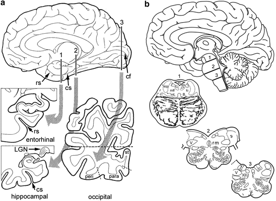
Brains obtained at autopsy—following precautions for handling tissue of prion diseases—are fixed by immersion in 10 % formalin (4 % aqueous solution of HCHO) for 1 week or longer. Remove the brainstem and cerebellum and cut the brain mid-sagittally. Take one hemisphere and remove the meninges to uncover the rhinal sulcus and the calcarine fissure. The staging procedure for AD requires evaluation of a few sections from a minimum of (A) two blocks of cortical tissue and (B) one brainstem block. The first block includes anteromedial portions of the temporal lobe (frontal section at the level of the mamillary bodies), encompassing uncal portions of the hippocampus, the parahippocampal gyrus, and adjoining temporal gyri (Braak et al. 2006a). The cutting line typically runs through the rhinal sulcus, including the transentorhinal and entorhinal regions (Fig. 11.1a, cutting line 1). The tissue on one side of the cut can be used for conventional paraffin embedding (70-100 ¢m sections; see Feldengut et al. 2013), and that on the other side for frozen sections or polyethylene glycol (PEG) embedding (100-400 μm sections). This block of temporal lobe tissue is essential for assessment of stages 1a, 1b, and NFT stages I–IV, and it contains the transentorhinal and entorhinal regions. A section thickness of 70-100 μm meets the demands of low power light (stereo)microscopy and greatly facilitates recognition of the lesional distribution pattern (Fig. 11.1)
.
Fig. 11.1
Tissue blocks recommended for neuropathological staging of AD. Abbreviations: aq—mesencephalic queduct; cf—calcarine fissure; cs—collateral sulcus; dtG—dorsal tegmental nucleus of Gudden; io—inferior olivary complex; lc—locus coeruleus; LGN—lateral geniculate nucleus; ml—medial lemniscus; mlf—medial longitudinal fascicle; para—parastriate area; peri—peristriate area; rm—great raphe nucleus; rs—rhinal sulcus; scp—decussation of superior cerebellar peduncle; st—striate area; V—motor nucleus of the trigeminal nerve; X—dorsal motor nucleus of the vagal nerve; XII—motor nucleus of the hypoglossal nerve (Adapted with permission from H Braak and E Braak, Exp Neurol 1998;153:351–365)
The conventional block of hippocampal tissue usually is removed at the level of the lateral geniculate nucleus (Fig. 11.1a, cutting line 2). However, at this latitude the adjoining parahippocampal gyrus often includes only posterior remnants of the transentorhinal and entorhinal regions. Since evaluation of these regions is essential to the staging procedure, care should be taken to remove the temporal block a few millimeters anterior to the level of the lateral geniculate nucleus (Braak et al. 2006a).
The second block needed for staging is removed preferably halfway between the occipital pole and the junction of the parieto-occipital sulcus with the calcarine fissure (Fig. 11.1a, cutting line 3). The cut is oriented perpendicular to the calcarine fissure. The size of the block can be reduced along the dashed line to fit conventional cassettes but should include the cortex covering the lower bank of the calcarine fissure and the adjoining basal occipital gyri. It shows occipital association areas, the parastriate area, and an easily delineable primary field, the striate area. This occipital block is essential for recognition of neurofibrillary stages V–VI.
The brainstem block needed for the diagnosis of the tau process is a section through the locus coeruleus and dorsal raphe nucleus at about the level 1 in Fig. 11.1b. The other levels are required for additional diagnoses of neurodegenerative diseases other than AD.
Specific antibodies for the pathologic proteins are available. However, their application often requires special fixation methods and/or antigen retrieval methods which influence the result of the immunoreaction to a variable extent. Dye staining techniques (congo red, thioflavine S) are rather insensitive, while conventional silver methods show both normal and pathological components (Bielschowsky, Bodian). This may be advantageous in some instances but usually impedes differentiation of the changed components from uninvolved parts of the brain.
Related posts:
 Basic Organization of Non-thalamic Nuclei with Diffuse Cortical Projections
Microtubules and the Protein Tau
Early Presymptomatic Stages
The Pattern of Cortical Lesions in Preclinical Stages
Introduction
The Pattern of Lesions During the Transition to the Symptomatic Phase and in Fully Developed Alzheimer’s Disease
Basic Organization of Non-thalamic Nuclei with Diffuse Cortical Projections
Microtubules and the Protein Tau
Early Presymptomatic Stages
The Pattern of Cortical Lesions in Preclinical Stages
Introduction
The Pattern of Lesions During the Transition to the Symptomatic Phase and in Fully Developed Alzheimer’s Disease
Stay updated, free articles. Join our Telegram channel

Full access? Get Clinical Tree


