Figure 2-1 A, Axial T1-weighted fast spin echo (FSE) magnetic resonance imaging scan obtained at 3T of the distal arm just above the level shows marked enlargement of the median nerve (arrows). The ulnar nerve (arrowhead) is normal. B, T2-weighted FSE image with fat suppression at the same level as A shows marked enlargement and T2 hyperintensity in the individual fascicles of the median nerve (arrows). C, T1-weighted spoiled gradient recalled echo (SPGR) image with fat suppression at the same level as A and B shows no enhancement (arrows) of the nerve, which is consistent with an inflammatory neuropathy.
Case B
This 12-year-old child presented with progressive, painless weakness in her left hand over a number of years. An MRI performed at another institution showed an enhancing mass. EMG indicated a nonspecific brachial plexitis. MRI of the left brachial plexus was performed with a 3T imager prior to and after contrast administration. The examination showed enlargement and T2 hyperintensity (Fig. 2-2A) of the entire brachial plexus, most prominently involving the posterior cord. After contrast there was avid enhancement of the enlarged, individual fascicles (see Fig. 2-2B). This is different from the lack of enhancement seen in the patient in Case A. This appearance is very characteristic of perineurioma, particularly when accompanied by muscle atrophy, as was the case here. The posterior cord was biopsied using targeted fascicular nerve biopsy and the final diagnosis was perineurioma. No treatment with steroids or intravenous immunoglobulin was warranted.
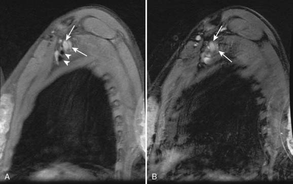
Figure 2-2 A, Sagittal T2-weighted fast spin echo (FSE) magnetic resonance imaging scan with fat suppression obtained at 3T of the brachial plexus shows marked enlargement and T2 hyperintensity in the medial and posterior cords of the brachial plexus (arrows). The adjacent axillary artery and vein are normal (arrowheads). B, Sagittal T1-weighted spoiled gradient recalled echo (SPGR) with fat suppression at the same level as A shows avid contrast enhancement of the affected portions of the plexus (arrows).
Related posts:
 Hot and Cold Feet—Sensory Neuropathy Associated with Human Immunodeficiency Virus
Borrelia Neuropathy with Necrotizing Vasculitis
Adrenomyeloneuropathy Masquerading as Charcot-Marie-Tooth Disease
Focal Mononeuropathy Onset of Amyotrophic Lateral Sclerosis
A Case of Guillain-Barré Syndrome Associated with Anti-GD1b Immunoglobulin G Antibodies
Length-Related Axonal Loss in Neuropathy
Hot and Cold Feet—Sensory Neuropathy Associated with Human Immunodeficiency Virus
Borrelia Neuropathy with Necrotizing Vasculitis
Adrenomyeloneuropathy Masquerading as Charcot-Marie-Tooth Disease
Focal Mononeuropathy Onset of Amyotrophic Lateral Sclerosis
A Case of Guillain-Barré Syndrome Associated with Anti-GD1b Immunoglobulin G Antibodies
Length-Related Axonal Loss in Neuropathy
Stay updated, free articles. Join our Telegram channel

Full access? Get Clinical Tree


