Fig. 8.1
Sagittal T1-weighted image without contrast. (1) clivus; (2) pons; (3) third ventricle; (4) sphenoid sinus; (a) anterior pituitary gland; (p) posterior pituitary gland; asterisk indicates pituitary stalk

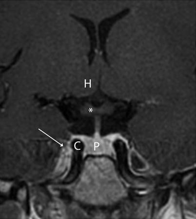
Fig. 8.2
Coronal T1-weighted image without contrast. (P) pituitary gland. (C) cavernous sinus; (H) hypothalamus; asterisk indicates optic chiasm
In men, the height of the pituitary gland remains somewhat constant following puberty. In women, the peak height of the normal pituitary gland occurs in the second to third decade of life, and the height of the gland may decrease thereafter [3].
On computed tomographic (CT) imaging, the normal pituitary gland is isodense to the brain and may appear homogeneous or heterogeneous [4]. Following magnetic resonance imaging (MRI) contrast administration, the normal gland usually enhances brightly.
Over 90 % of normal pituitary glands have flat or concave superior surfaces along the diaphragm sella [5]. The pituitary stalk (infundibulum) connects the gland to the hypothalamus and runs through the diaphragm sella; it should be seen on the midline.
The neurohypophysis appears as a “bright spot” on standard T1-weighted MRI in over 90 % of healthy participants [6].
8.1.1 Sellar and Sphenoid Sinus Anatomy on MRI
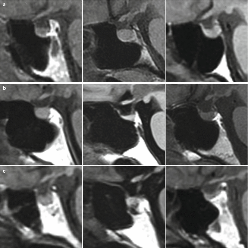
Three general morphologies of the sellar floor have been described on sagittal MRI (Fig. 8.3) [7]:

Prominent (sellar angle of <90°) in 25 % of people
Curved (sellar angle 90–150°) in 63 % of people
Flat (sellar angle >150°) in 11 % of people
No floor (conchal sphenoid) in 1 % of people

Fig. 8.3
Normal variants of sellar and pituitary morphology seen on sagittal post-contrast MRIs. (a) The top row depicts prominent sellar floor morphology that is easy to identify. (b) The middle row shows normal variants with a curved, intermediate morphology. (c) The bottom row shows examples of a “flat” sella with a partially pneumatized sphenoid sinus that is harder to identify intraoperatively (Adapted from Zada et al. [7]; with permission)
In healthy adults, mean measurements on sagittal MRI of the following structures are as follows: sellar face length, 13.4 mm; sellar prominence, 3.0 mm; sellar angle, 112°; angle of tuberculum sellae, 112°; and sellar-clival angle, 117°. The average width of the normol sella is 12.7 mm [7].
A simple sphenoid sinus configuration, defined as having no septa, one vertical septum, or two symmetric vertical septa, is noted in 71 % of studies. In the other 29 % of people, an MRI shows a complex configuration, defined as two or more asymmetric septa, three or more septa of any kind, or the presence of a horizontal septum [7].
On MRI, the normal distance between the internal carotid arteries (ICAs) at the parasellar and midclivus levels are 16.2 and 18.5 mm, respectively [7].
Stay updated, free articles. Join our Telegram channel

Full access? Get Clinical Tree


