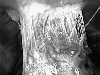
17 Diagnosing a thoracic vertebral lesion can be a formidable task. The traditional forms of biopsy include open excisional or incisional biopsy and needle or aspiration biopsy. Open biopsy requires a major surgical procedure and exposes the patient to the risks of a thoracotomy. Needle biopsy using computer tomographic (CT) guidance has been shown to be safe and reliable.1 However, many radiologists are reluctant to perform a CT-guided biopsy high in the thoracic spine, especially one located near the spinal cord or aortic arch. Percutaneous passage of a needle in the thoracic spine also runs the risk of creating a pneumothorax.2 Additionally, needle biopsies are prone to sampling error. They yield only small amounts of tissue and therefore may not result in a definitive diagnosis. In cases of presumed infection and well-defined lesions, needle biopsy offers a reasonably noninvasive and effective tool to reach a diagnosis. For more complex lesions, however, needle biopsy may be inadequate. Thoracoscopic biopsy can provide a minimally invasive alternative for the diagnosis of spinal and paraspinal lesions. Thoracoscopic biopsy can minimize a patient’s discomfort and obviate the need for an open procedure, eliminating the associated risks, including spinal instability. Compared with percutaneous techniques, thoracoscopy also offers obvious advantages: direct visualization of the biopsy site, avoidance of vital structures, adequate sample size, and ease of hemostasis after a biopsy is obtained. The historical roots of thoracoscopy lie in the development of early 19th century devices for visualizing body cavities. As recounted elsewhere,3 the first such device for medical practice, used in 1806 by Philippi Bozzini, was created by attaching a candle to a thin cannula. This endoscopic device of sorts was known as a lichtleiter3 and possessed no optics. In 1853, however, Desormeaux introduced the use of a lens to focus the light source.3 After optics were introduced to these first endoscopes, Bevan undertook the first esophagoscopy in 1868. The next major advance occurred in 1870, when Kussmaul performed the first esophagogastroscopy by adding a more powerful light source.3 The cystoscope was then introduced in 1879. In addition to a powerful light source and optics, Nitze’s “Blasenspiegel” (bladder mirror) featured a working channel to allow both therapeutic and diagnostic procedures. Further refinements allowed the endoscope to be used to visualize the abdomen and pelvis in dogs in 1902 and in humans in 1929, respectively. The development of thoracoscopy rapidly followed the development of laparoscopy. In 1910 Jacobeus introduced the techniques of pleuroscopy and thoracoscopy.3 During the next two decades, thoracoscopy was widely accepted and performed in Europe; however, thoracoscopy was not popular in the United States until the 1970s, after several technical advances, including the use of fiberoptic technology and flexible operating scopes, were introduced.3 Later, video technology allowed surgeons to use both hands. This refinement led to the development of video-assisted thoracoscopic surgery (VATS).3 In the early 1990s, thoracoscopic spinal surgery was developed by two groups simultaneously. 3 Early techniques were limited to resection and biopsy of spinal and paraspinal lesions. The advent of minimally invasive spinal surgery has resulted in the adaptation of thoracoscopic techniques to even more complex procedures. In the next 100 years, the field should advance even further than it has since the invention of Bozzini’s lichtleiter. Compared with open thoracotomy, thoracoscopy has several advantages in terms of procedural ease and postoperative morbidity.4–10 Thoracoscopic procedures of the spine afford excellent visualization of spinal anatomy, perhaps even surpassing the visualization obtained during open thoracotomy.6 With open thoracotomy, the major source of pain and complications is related to spreading the ribs.6 This maneuver is minimized in thoracoscopic procedures. Consequently, the procedure is associated with much less trauma to the thorax and its contents, less postoperative pain, lower complication rates, and shortened hospital stays compared with open procedures.3 There are also several disadvantages to the use of thoracoscopy. First, the procedure cannot be used in patients with pleural adhesions caused by severe pulmonary disease. Adhesions prevent safe passage of the ports necessary to perform the procedure. Second, thoracoscopy does not allow access to the posterior aspect of the spine or to the contralateral pedicle. Finally, the skills required to be an effective and safe thoracoscopic surgeon are acquired only after progressing through a steep learning curve; however, recent improvements in instrumentation, optics, and technique training have improved the safety and efficacy of this technique dramatically.3,6,10,11 The principal reason to consider thoracoscopy over thoracotomy for the biopsy of spinal and paraspinal lesions is to avoid complications associated with the open procedure: pulmonary dysfunction, infection, prolonged pneumothorax, and postthoracotomy pain syndromes.9 Depending on the experience of the spine surgeon, thoracotomy also may require the presence of a cardiothoracic surgeon for the exposure and closure. Both thoracotomy and thoracoscopy provide an anterolateral approach to the spine with a full and direct view of the ventral surface of the spine.9,11 Incision size for a thoracotomy ranges from ~6 inches to as long as 15 inches; in contrast, thoracoscopy requires three to four 1-inch incisions.9,11 Thoracotomy also requires extensive transection of muscle, and 6 to 12 inches of rib must be resected or retracted. Thoracoscopy requires minimal muscle transection, and only about 1 inch of the head and proximal portion of the rib must be removed. No rib retraction is required.11 Therefore, thoracoscopy offers the same operative exposure as thoracotomy with smaller incisions and minimal muscular and bony resection, all of which help to reduce the morbidity rates associated with this procedure. Besides establishing a diagnosis, the indications for a thoracoscopic biopsy include seriously ill patients whose medical condition may preclude a more extensive open procedure and high thoracic lesions (especially T2–T4 on the left side). Lesions amenable to needle biopsy but of concern because of their high vascularity also can be approached thoracoscopically. Bleeding sites can be visualized directly and coagulated. Thoracoscopic biopsy is most useful when less invasive diagnostic studies fail to yield a definitive diagnosis. The two pathologic entities most frequently identified by thoracoscopic biopsy are malignancies and infections (especially vertebral osteomyelitis and discitis). Neoplastic lesions in the thoracic spine can be divided into primary (benign/malignant) or metastatic categories. When a noninvasive evaluation fails to yield a diagnosis, thoracoscopic biopsy not only helps establish a diagnosis but also helps guide future therapy. This option can be especially important when radiation therapy and chemotherapy, rather than surgery, are the first line of treatment (e.g., plasmacytomas, Ewing’s sarcomas, lymphomas, hemangiomas, aneurysmal bone cysts, and eosinophilic granulomas). Metastatic lesions are much more common than primary malignancies of the spine. If a primary diagnosis cannot be established, these lesions can be biopsied to help institute treatment and to help stage the extent of disease spread. Finally, patients too ill to tolerate a major procedure or with a poor functional status are ideal candidates for the minimally invasive thoracoscopic approach. Infections of the spine may be subdivided into discitis and osteomyelitis. Definitive identification of the offending organism is required to help tailor antimicrobial therapy, particularly with the increase in multidrug-resistant organisms. Thoracoscopic biopsy not only can be used for diagnosis, but also can be used as an adjuvant to antibiotic therapy. Localized fluid collections can be drained, and antibiotics can be irrigated locally with thoracoscopy. In the case of certain fastidious organisms, the large quantity of tissue obtainable by thoracoscopic biopsy may be the only way to establish the causative organism without an open procedure. In general, contraindications to thoracoscopy are limited to those associated with the approach. For the hemithorax to be entered safely, a patient must be able to tolerate a general anesthetic. Patients also must be able to tolerate single-lung ventilation because the ipsilateral lung must be collapsed during the procedure. Additional contraindications can include a previous thoracotomy or a history of empyema on the ipsilateral side. Both conditions can lead to pleural adhesions, which limit the surgeon’s ability to work in the pleural space with thoracoscopic instruments. Contraindications to thoracoscopic spinal surgery fall into two categories: airway-pulmonary and spinal. Airwaypulmonary contraindications include age less than 5 years, tracheobronchial stenosis, prior tracheostomy, tracheobronchial neoplasm, tracheobronchial scar tissue, congenital stenosis, or aplasia.12 The procedure itself requires patients to undergo single-lung ventilation; therefore, any pulmonary pathology that would preclude single-lung ventilation would be a contraindication to thoracoscopy: pulmonary edema, acute respiratory distress syndrome, pneumonia, acute congestive heart failure, lung neoplasm (primary and metastatic), chronic obstructive pulmonary disease, severe reactive airway disease or asthma, interstitial pulmonary fibrosis or pneumonitis, pulmonary hypertension, acute or chronic respiratory insufficiency, contralateral bronchopleural fistula, contralateral pulmonary artery aplasia, contralateral pneumothorax, contralateral lobectomy or pneumonectomy, contralateral lung aplasia, congenitally anomalous tracheobronchial tree, paralytic hemidiaphragm, contralateral pleural effusion, or empyema.12 Spinal contraindications are relative and include intradural extension of the pathology to be biopsied, pathology affecting the posterior and contralateral elements of the spine, prior cerebrospinal fluid (CSF) leakage, prior surgery at the biopsy site, a highly vascular tumor, and vascular anomalies.12 Once the patient is placed in the lateral decubitus position with the hemithorax to be explored facing upward, the primary portal is placed in the posterior axillary line at the same level as the lesion. The endoscope is inserted through this portal. Typically, the first portal is placed rostral to the sixth intercostal space to avoid injury to the diaphragm. After a 1-cm long incision is made, blunt dissection with a Kelly clamp or hemostat allows entry into the pleural space. Before the trocar is placed, blunt finger dissection can be used to release any adhesions. Once the endoscope is in position, the remainder of the portals can be placed under direct visualization. Triangulation is the goal of portal placement. This configuration allows the surgeon to work from above and below the intended target. Each access portal is placed in the anterior axillary line, and additional working portals may be placed as needed to complete the case. The order of portal placement depends on the level of the lesion being biopsied. For lesions high in the thoracic spine, the initial (posterior) portal may be placed high (i.e., the fourth intercostal space) in the posterior axillary line. For lesions low in the thorax, the T6 portal remains standard to ensure the safety of the diaphragm and intraabdominal compartment. The next portal is placed just above the diaphragmatic insertion in the anterior axillary line. It allows a retractor to be placed to mobilize the diaphragm inferiorly. Once the diaphragm is retracted inferiorly, lower working portals are placed under direct visualization. After the portals are placed, the thoracic compartment is inspected and the vital structures within the thoracic compartment are identified (Fig. 17-1). Once oriented, the surgeon’s attention should be directed at the lesion in question. The lesion is localized by inspection and then confirmed radiographically (Fig. 17-2). The surgeon counts the ribs from within the chest cavity to confirm the level of the lesion. Typically, a fat pad at the apex of the thorax hides the first rib; therefore, counting begins with the first visualized rib, T2, and proceeds caudally. Once the lesion has been found, a spinal needle is placed into the disc space above (or below) the lesion. Cross-table anteroposterior radiography is obtained to confirm the location of the lesion before biopsy.
Thoracoscopic Biopsy of Spinal and Paraspinal Lesions
 History of Thoracoscopy
 Advantages and Disadvantages of Thoracoscopy
 Thoracoscopy Versus Thoracotomy
 Indications
Thoracoscopic Diagnosis of Tumors
Thoracoscopic Biopsy for Infection
 Contraindications
 Surgical Technique
Related posts:
 Lumbosacral and Pelvic Reconstruction
 Anatomy of the Spine and Spinal Cord
Stay updated, free articles. Join our Telegram channel

Full access? Get Clinical Tree


