Fig. 6.1
A supraorbital flap (dark black), based on the true keyhole, may be modified by involving more of the lateral orbit, extending medially (bifrontal) or extending temporally with or without a zygomatic osteotomy
6.3 Frontal Approaches
The unifrontal or bifrontal approach is the oldest cranial approach employed for lesions of the sellar region [5]; its main advantage is its “straight-shot” trajectory beneath the frontal lobe to the optic chiasm and sella. A lumbar drain facilitates brain relaxation. A three-fourths bicoronal incision or a full bicoronal incision, sparing the temporalis, is fashioned behind the hairline for a unilateral or a bilateral subfrontal approach, respectively. Excessive extension of the head results in an overly oblique orientation of the planum and tuberculum away from the surgeon. A pericranial flap from one temporal line to the other, based anteriorly on the supraorbital vessels and extending under the posterior aspect of the incision, should be raised during the opening in anticipation of frontal sinus entry or violation of the ethmoid or sphenoid sinuses, which would merit repair with a vascularized graft. A bifrontal craniotomy follows, which should be taken as low as possible to decrease the distance to the anterior fossa floor and minimize brain retraction.
The dura is opened low and transversely—across a ligated sagittal sinus, if bilateral—and is reflected inferiorly. The olfactory tracts are identified and separated from the frontal lobe to prevent avulsion. The optic nerves and chiasm are readily seen with gentle frontal lobe retraction. Ventricular extension of the lesion may be reached through the lamina terminalis; limitations include a prefixed optic chiasm and lateral/cavernous sinus or substantial superior extension. Additional extradural dissection after frontal craniotomy may add to the exposure in select cases; the planum and tuberculum may be drilled for more inferior exposure beneath a prefixed chiasm, and the optic canals can be opened for greater maneuverability around the optic apparatus. Any parasellar or frontal sinus should be exenterated, obliterated, and covered with a large pericranial graft.
The surgeon may also consider the subfrontal “keyhole” approach [6], through either an eyebrow or eyelid incision. This approach has been used to access the sellar and parasellar regions. The incision most commonly is placed in the eyebrow with a slight lateral extension, being sure to stay lateral to the supraorbital nerve. The craniotomy, intended to be situated lateral to the frontal sinus, is small, and the main advantage is cosmetic.
6.4 Supraorbital Approach
The supraorbital approach, like the bifrontal approach, provides access to pathology of the planum, tuberculum, clinoid, and sella with minimal or no extension lateral to the carotid. The main conceptual advantage is that removal of the superior and lateral orbital rims reduces the amount of frontal lobe retraction required and may facilitate removal of more superiorly projecting midline tumors through enhanced visualization.
We perform the supraorbital approach based on the approach originally described by Jane et al. [7]. A spinal drain is optional. The scalp incision is begun 1 cm anterior to the tragus and is continued in curvilinear fashion behind the hairline to the level of the superior temporal line on the opposite side. The superficial temporal artery is behind the incision, and the facial nerve branches are in front of it. A large pericranial flap based on the supraorbital and frontal vessels, beginning well behind the skin incision and extending from one superior temporal line to the other, is raised and reflected over the scalp flap anteriorly and dissected off the superior and lateral orbital walls. The supraorbital nerve is freed from its notch or freed from its foramen with a high-speed drill.
Only the superior aspect of the temporalis muscle needs to be mobilized. To do this, a subfascial dissection of the temporalis fascia is carried out by incising both layers of the fascia to expose the temporalis muscle, beginning at the keyhole and proceeding behind the course of the facial nerve to approximately the level of the sylvian fissure. The fascial layers are reflected forward, and the temporalis muscle, dissected free from its overlying fascia, is released from its insertion at the superior temporal line and along the superior and lateral orbit and reflected inferiorly. The junction of the zygomatic, sphenoidal, and frontal bones is thus exposed.
The bone flap is usually a unilateral supraorbital bone flap (Fig. 6.2a), although large tumors may be approached with a bifrontal supraorbital flap, with a one-piece intent for cosmetic purposes. The unilateral flap begins with placement of a burr hole at the anatomical keyhole about 1 cm behind the frontozygomatic suture. When drilled correctly, the superior half exposes frontal lobe dura and the lower half exposes the periorbita, with the orbital roof in the center of the burr hole separating the frontal lobe and orbit. An additional cut is made in the lateral orbital rim anywhere above the zygoma while the lateral periorbita is protected and continued to the orbital half of the keyhole. Finally, the flap is completed with a superior orbital roof osteotomy, done by placing a chisel straddling the lateral aspect of the superior orbit exposed in the keyhole and completing the osteotomy, with cottonoids protecting the frontal dura and the periorbita. It is important to angle the chisel superiorly, to ensure that the medial end of the osteotomy is well away from the orbital apex. It is also critical to complete the osteotomy rather than to “crack” the bone flap off manually, which may lead to unintended angles of roof compromise through the optic canal.
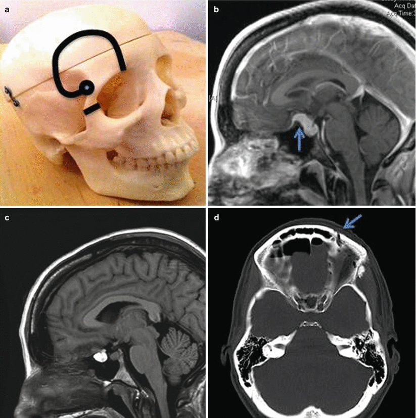
Fig. 6.2
(a) Supraorbital bone flap. (b) Sagittal T1 contrast MRI showing a tuberculum sella meningioma with significant hyperostosis (blue arrow). (c) Postoperative T1 MRI showing fat in the roof of the sphenoid sinus after involved bone is drilled away. (d) Postoperative CT scan showing the superior orbital osteotomy just lateral to the frontal sinus (arrow)
If the frontal sinus is entered during the exposure, the sinus is exenterated and its posterior wall is removed. A small piece of fat or temporalis muscle is used to pack the sinus and obliterate the frontonasal duct.
This approach provides excellent access to the optic nerves and chiasm and to pathology in the sellar region in or in front of the sella and medial to the carotids. Both optic canals can be accessed and opened from this vantage point.
The pituitary stalk is distinct in its color and vascular arborization. Tuberculum sellae meningiomas usually displace the stalk posteriorly; rarely, they will engulf the stalk, requiring careful microdissection. Hypophyseal branches off the carotid are spared.
The involved dura of the tuberculum and planum is resected and any hyperostotic bone is drilled with a high-speed diamond burr. Paranasal sinus entry is handled by exenterating the sinus mucosa and packing the spaces with fat. A piece of free pericranium or fascia lata is then placed intradurally and secured to the native dura over the resected dura. After dural closure, the pericranial flap is turned over the orbit and frontal bone and is spread over any defect in the anterior fossa floor.
6.4.1 Application to Parasellar Tumors: Tuberculum Sellae Meningiomas
Tuberculum sellae meningiomas, originating in front of the sella and arising from the tuberculum, chiasmatic sulcus, and limbus sphenoidale, accounted for 5–10 % of meningiomas in Cushing’s and Eisenhardt’s series [8]. These tumors often invade the floor of the anterior fossa and cause hyperostosis, commonly in front of the sella (Fig. 6.2b). As they enlarge, they displace the optic nerves laterally and the chiasm superiorly; they also impinge on the pituitary stalk posteriorly, as well as the basilar artery, and they can extend to the interpeduncular space. They frequently insinuate into one or both optic canals. The blood supply is typically inferiorly from the posterior ethmoidal arteries. Meticulous attention must be paid to the anterior cerebral artery (ACA) complex, which (depending on the tumor’s size) may be encased or displaced superiorly. Careful attention also must be paid to small, medial lenticulostriates.
6.4.2 Specific Surgical Principles
The main principles of the supraorbital approach as it pertains to tuberculum sellae tumors are the low frontobasal exposure, which facilitates access to the tumor without undue frontal retraction, and access to both optic canals (Fig. 6.2c, d). Once the dura is opened, the ipsilateral olfactory tract is quickly encountered and should be dissected off from the frontal lobe in order to prevent its avulsion. A drop of fibrin glue applied to the olfactory tract may further decrease the risk of damage to the tract. When the tumor is encountered, its basal posterior ethmoidal arterial feeders are coagulated to devascularize the tumor; central debulking then may be undertaken by ultrasonic aspiration or other means. The optic chiasm is usually displaced superiorly, and the optic nerves, superiorly and laterally. Should the nerves be engulfed in tumor, dissection should begin at the chiasm and proceed proximally. The arterial supply to the optic chiasm and nerves is preserved. All associated dura is resected, and any hyperostotic bone at the tuberculum and planum is drilled away with a high-speed diamond bit.
Decompressing the visual apparatus is not complete until the optic canals are unroofed. In a recent series, 67 % of tuberculum sellae tumors were associated with tumor extension into one optic canal (40 %) or both canals (60 %) [9]. Moreover, tumor left inside the optic canal may be the source of recurrence or of diminished visual improvement after surgery [10]. One or both optic canals are exposed and unroofed with a high-speed drill under constant irrigation, to avoid thermal injury to the optic nerve. The falciform ligament and optic sheath are opened sharply. Optic nerve decompression should proceed as far anterior toward the globe as is needed to ensure that all optic extension of the tumor has been extirpated. Even if the clinical presentation suggests extension of the tumor into only one optic canal, the surgeon should be prepared to decompress both canals on the basis of intraoperative findings. Even the slightest suggestion of canalicular extension should prompt at least the opening of the falciform ligament.
6.5 Pterional Approach
The pterional or frontotemporal approach, popularized by Yasargil and Fox [11], has the additional benefit of providing a lateral corridor to sellar lesions, access to cavernous sinus extension, and more superior access than afforded by an anteriorly directed frontal or supraorbital approach. Importantly, it allows for a greater degree of vascular control, given the excellent access it provides to the parasellar vasculature. Lesions that extend to the cavernous sinus should be approached with, at minimum, a pterional approach.
The placement of a lumbar drain is optional; it is less important than in frontal approaches because of the access the pterional approach affords to the sylvian cisterns. The head is turned 30° away and extended, bringing the lateral aspect of the malar eminence to an uppermost position. This position also usually places the ipsilateral optic nerve perpendicular to the floor.
Related posts:
 Granular Cell Tumors
 Arteriovenous Malformations
 Olfactory Neuroblastoma and Sellar Neuroblastoma
 Traumatic Injury of the Sellar Region, Pituitary Stalk Disruption, and Posttraumatic Anosmia
 Schwannomas of the Sellar and Parasellar Region
 Nasopharyngeal Carcinoma and Squamous Cell Carcinoma of the Paranasal Sinuses
Stay updated, free articles. Join our Telegram channel

Full access? Get Clinical Tree


