Transcranial Magnetic Stimulation (TMS): Clinical Applications


Transcranial Magnetic Stimulation (TMS): Clinical Applications


Alexander Rotenberg

Alvaro Pascual-Leone


Transcranial magnetic stimulation (TMS), a well-tolerated method for focal noninvasive brain stimulation, has emerged as a realistic clinical tool in neurology (1,2). As diagnostic procedures, single-pulse TMS and paired-pulse TMS may be used to noninvasively map cortical function and to measure regional cortical excitability (3,4). In therapeutic applications, the capacity of repetitive TMS (rTMS) to induce a lasting change in cortical excitability has been tested in several disease states, including epilepsy (5, 6, 7, 8 and 9). The 2008 United States Food and Drug Administration (FDA) clearance of the Neurostar device and rTMS protocol for treatment of some patients with medication-resistant major depression underscores its gain of acceptance in the clinical setting.


MEASURES OF MOTOR CORTEX EXCITABILITY BY TMS IN EPILEPSY

In most common protocols, TMS is coupled with surface EMG (TMS-EMG) such that the motor cortex is stimulated and the magnitude of the evoked muscle contraction in a contralateral limb (typically a hand muscle) can be quantified by skin electrodes and the recording of a motor evoked potential (MEP) (reviewed in Ref. 2). From the MEP, a number of measures can be derived to probe cortico-spinal excitability (Table 53.1) and a number of them characterize cortical and intracortical excitation/inhibition balance. One is the threshold to muscle activation or motor threshold (MT). The MT, obtained by single-pulse TMS, appears to reflect largely sodium channel-mediated membrane excitability in efferent pyramidal cells, and is increased by anticonvulsants, such as phenytoin and carbamazepine that inhibit voltage-gated sodium channels. Additionally, paired-pulse TMS (
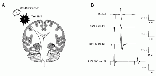
Fig. 53.1) provides measures of γ-aminobutyric acid (GABA)-mediated corticocortical inhibition and glutamate-dependent cortico-cortical excitability. In the most common paired-pulse TMS protocols, a subthreshold conditioning stimulus is delivered before each succeeding TMS pulse (reviewed in Refs. 2, 10). Short (1 to 5 msec) interstimulus intervals lead to reduction of the MEP, and likely reflect GABAA receptor-mediated short-interval intracortical inhibition (SICI). Slightly longer (6 to 20 msec) interstimulus intervals augment the MEP, reflecting glutamatemediated intracortical facilitation (ICF). Benzodiazepine (GABAA receptor agonist) anticonvulsants such as diazepam and lorazepam enhance SICI and suppress ICF (11, reviewed in Ref. 12). Still longer interstimulus (50 to 300 msec) paired-pulse TMS-EMG protocols can also measure GABAB receptor-mediated long-interval intracortical inhibition (LICI) that is enhanced by the GABAB receptor agonist baclofen (13,14). The extent of cortical inhibition may also be measured by the cortical silent period (CSP), a transient EMG silence observed when TMS is delivered to the motor cortex during an active motor contraction. The CSP too appears mediated by GABA receptors, although the contributions of GABAA and GABAB receptors to CSP is less defined than for paired-pulse measures (11,15,16).

These single-pulse TMS and paired-pulse TMS measures appear useful in detecting abnormalities in the inhibition:excitation ratio in patients with epilepsy. Data from published reports where parameters derived from TMS-EMG in patients with epilepsy were compared to values obtained from nonepileptic controls are summarized in Table 53.2. Although findings vary between studies, and likely reflect subject and methodology differences, they do suggest that either primary or compensatory abnormalities in cortical inhibition:excitation ratio can be measured by TMS. In particular, pathologic suppression of intracortical inhibition (Table 53.2) as detected by paired-pulse stimulation appears to be a common finding in patients with epilepsy.

Detection of abnormalities in cortical inhibition by TMS-EMG data suggests its possible utility in epilepsy, but also underscore a limitation as global cortical excitability must be inferred from stimulation of the motor cortex. However, this anatomic limitation of TMS methods that require simultaneous EEG may be overcome by TMS-EEG where the brain can be safely stimulated and TMS-evoked surface potentials can be recorded with scalp electrodes and used to estimate regional excitability of the extramotor cortex (34, 35 and 36).

In support of potential TMS-EEG application in measuring cortical excitability in patients with epilepsy, a recent experiment demonstrates that inhibition of the evoked EEG response over extramotor frontal and parietal cortex, analogous to LICI, can be recorded with a paired-pulse TMS-EEG paradigm (37,38). An interesting extension of these data may be to test whether extramotor LICI TMS-EEG abnormalities are present in the epilepsy population. However, as number of TMS-EMG experiments show motor cortex abnormalities in patients with extramotor and generalized epilepsies, further studies will be required to test whether interrogating focal cortical excitability outside of the motor cortex by TMS-EEG is of any greater clinical value than checking TMS-EMG measures (39, 40 and 41).


Table 53.1 TMS-EMG Parameter


TMS-EMG Parameter


Protocol


Likely Mechanism


Examples of Change with Medication


Motor threshold (MT)


Single-pulse stimulation: measure of stimulus strength necessary for a motor response


Corticomotor neuron membrane excitability


Increased by sodium channel antagonists (e.g., PHT, CBZ)


Short-interval intracortical inhibition (SICI)


Paired-pulse stimulation: conditioning stimulus precedes test stimulus by 1-5 msec


GABAA-medicated inhibition


Increased with GABAA agonists (LZP, DZP)


Intracortical facilitation (ICF)


Paired-pulse stimulation: conditioning stimulus precedes test stimulus by 6-20 msec


Glutamate-mediated excitation


Decreased with GABAA agonists (LZP, DZP)


Long-interval intracortical inhibition (LICI)


Paired-pulse stimulation: conditioning stimulus precedes test stimulus by 50-300 msec


GABAB-mediated inhibition


Increased by GABAB agonists (baclofen; PGB)


Cortical silent period (CSP)


Single-pulse stimulation: measure of pause in voluntary EMG activity after TMS


GABAB-mediated inhibition


Increased by GABAB agonists (baclofen; PGB); decreased by GABAA agonists (LZP, DZP)


CBZ, carbamazepine; DZP, diazepam; LZP, lorazepam; PGB, pregabalin; PHT, phenytoin.


From Rotenberg A. Prospects for Clinical Applications of Transcranial Magnetic Stimulation and Real-Time EEG in Epilepsy. Springer Science+Business Media, LLC; 2009.


TMS IN PRESURGICAL FUNCTIONAL MAPPING

rTMS has been applied to lateralize expressive language, and thus may have a role in epilepsy surgical planning. Although language lateralization through rTMS-induced speech arrest shows a fairly high concordance with the results of intracarotid amytal (Wada) testing in epilepsy patients (42, 43 and 44), there are some drawbacks for cortical mapping of linguistic functions through this approach. Online rTMS appears to lack sensitivity for determination of language dominance, as some studies report difficulties to obtain speech arrest in more than one third of all tested patients (43,45). Even when rTMS parameters are adjusted to reliably induce speech arrest, online rTMS shows a relatively poor prognostic value for postoperative language deficits. As compared to the Wada test, the results of rTMS-induced speech arrest do more often favor the right hemisphere and match less often the postoperative outcome with respect to language deficits (46). This is most likely accounted for by the fact that speech arrest is obtained most easily over facial motor areas, where true aphasia is rarely observed (47). Speech arrest might thus not represent an optimal marker for language lateralization. In future studies, rTMS protocols will have to be adapted in order to target other aspects of language than speech production, if online rTMS is to become a useful tool in presurgical evaluation of epileptic patients (46). Of special interest in this respect is a study on the susceptibility of Wernicke’s area to rTMS-induced language disruption (in a picture-word matching task) and the relationship to language lateralization in the same healthy subjects as assessed through fMRI (48). This study showed that rTMS-induced language disruption is significantly affected by the degree of hemispheric language lateralization. Whether this or other protocols might be useful in patients with epilepsy for a precise determination of language dominance awaits future investigations.


Figure 53.1 Paired-pulse TMS illustration. A: Schematic shows a paired-pulse protocol where two successive stimuli are delivered unilaterally to the motor cortex. B: Representative motor evoked potentials (MEPs) modulated in size as a function of the interstimulus interval (ISI) are shown. Relative to the control stimulus, a short (2 msec) ISI results in short-interval intracortical inhibition (SICI) of the test MEP, slightly longer (12 msec) ISI leads to intracortical facilitation (ICF), and still longer ISI (200 msec) produces long-interval intracortical inhibition (LICI) of the test MEP.


Table 53.2 TMS-EMG Findings in Patients with Epilepsy


Findings (Relative to Control); Comments


Study


Subjects (n)


MT


SICI


ICF


LICI


CSP


Akgun et al. (17)


JME(21); AS(21); C(20)


↑; all on AED


NT


NT


NT


Badawy et al. (18)


JME(10); GE(10); FE(10); C(10)


=


↓/=; reduced in early morning in JME, but not other GE/normal in FE


↓; reduced in early morning in all GE; normal in FE


NT


Brodtmann et al. (21)


JME(3); GTC(3); JAE(1); C(16)


=


NT


NT


NT


Cantello et al. (20)


FE(18); C(11)


↑; all on AED


↓; reduced in a subject of patients with FE


↑; increased in a subset of patients with FE


NT


NT


Caramia et al. (21)


JME(7); GTC(2); C(6*) *1 nonepileptic subject on PB


↑/=; increased in JME/normal in GTC; all on AED


↓; reduced in JME


NT


NT


NT


Cicinelli et al. (22)


FE(16); C(16)


NT


NT


NT


↓; reduced in hemisphere ipsilateral to seizure focus


Cincotta et al. (23)


FE(18); C(16)


↑; all AED


NT


NT


NT


↑; increased in subgroup with clonic seizures


Danner et al. (24)


EPMI(24); C(24)


↑; majority on AED


NT


NT


NT


Di Lazzaro et al. (25)


E(5); C(11)


↑; all on AED


=; increased with VNS


NT


NT


NT


Ertas et al. (26)


GE(14); FE(11); C(14)


NT


NT


NT


NT


Fedi et al. (16)


GE(14, all with GABRG2(R43Q) mutation); C(24)


=


NT


=


Hamer et al. (27)


FE(23); C(20)


↑; majority on AED


=


=


NT


=; reduced in hemisphere ipsilateral to extramotor seizure focus


Klimpe et al. (28)


GE(10): FE(10); C(20)


=


=


NT


=


Macdonell et al. (29)


JME(9); JAE(1); GE(11); C(19)


=


NT


NR


NT


Manganotti et al. (30)


JME(15); C(12)


=


=


=


=


Manganotti et al. (31)


JME(10); C(10)


=


↓; reduced further with sleep deprivation


=


NT


=


Manganotti et al. (32)


JME(9); C(20)


=


=


NT


NT


Molnar et al. (33)


FE(3); SGE(2); C(9)


↑; all on AED


=


↓; at 50 msec ISI


=


From Rotenberg A. Prospects for Clinical Applications of Transcranial Magnetic Stimulation and Real-Time EEG in Epilepsy. Springer Science+Business Media, LLC; 2009.

Only gold members can continue reading. Log In or Register to continue

Stay updated, free articles. Join our Telegram channel

Sep 9, 2016 | Posted by in NEUROSURGERY | Comments Off on Transcranial Magnetic Stimulation (TMS): Clinical Applications

Full access? Get Clinical Tree

Get Clinical Tree app for offline access