Fig. 32.1
This figure shows the differences visible on lateral radiographs in the size proportions within the lumbar spine in a 20-year-old adult (a), in a growing spine at the age of 5 (b) and in a 4-year-old child with spondyloepiphyseal dysplasia (c). The most important parameters to measure when selecting the appropriate type of posterior osteotomy are the vertebral body height (red), the sagittal diameter of the vertebral body (yellow), and the thickness/height of the intervertebral disk (blue). Note that in a syndromic spine deformity, substantial variations of size dimensions are possible which necessitates individual planning by measuring above parameters at the site of the osteotomy

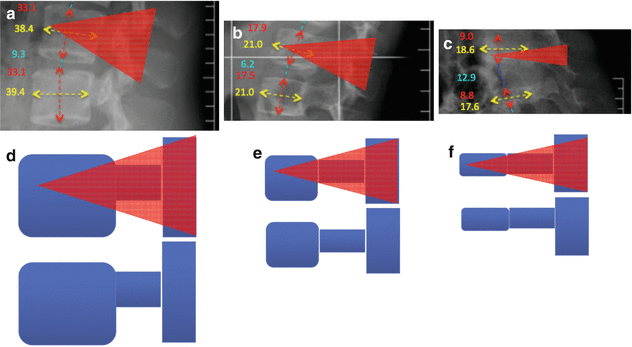
Fig. 32.2
The figure indicates the same parameters as in Fig. 32.1 as an example as measured in mm at the level of L2. The radiographs with illustrations (a–c) and illustrations (d–f) indicate what correction could be achieved by applying a PSO as indicated by the red triangle. Note that the spinal canal size in adults is almost reached at the age of 5 years, which means that the proportion in size of the anteroposterior diameter of the vertebral body and that of the spinal canal is smaller in children (d vs. e and f). These abovementioned size relations explain why less correction can be achieved when performing a PSO in children or patients with skeletal dysplasia patients compared to adults. Furthermore, a PSO in a child (b, e and c, f) would mean almost a vertebral resection. The consequences are twofold: the less competent anterior column (high disc space, low vertebral body) would then be even further weakened and one would loss an important anchoring point by removing the pedicles (no pedicle screws are possible at that level). Analyzing the size proportions becomes evident that a simple posterior SP-type osteotomy might be powerful enough to achieve sufficient correction in the growing spine
The pedicle subtraction osteotomy (PSO) can achieve in adults approximately 30° of correction in the sagittal plane at each spinal level at which it is performed. It is, however, rarely necessary in skeletally immature patients.
Any posterior osteotomy, be it SPO, PSO, or posterior vertebral column resection (PVCR), can also be performed in an asymmetrical way, to allow coronal plane correction.
A spinal deformity is always associated with a shortening of the spine. Conventional osteotomy techniques (some of the SPOs, PSO, VCR, hemivertebra resection) are coupled with resection and compression and thereby further shorten the spine.
In the growing spine, distraction seems to be a more logical approach to increase (normalize) the length of the spine, although intuitively it is expected to be associated with higher risks. Nonetheless, for congenital deformities with a concave side bar, an opening wedge osteotomy can be performed. It is not necessarily coupled with fusion of the osteotomized region.
Neurological injuries are the most feared complications following these procedures. There is an increased risk of neurological complications, compared with correction without any type of release or osteotomy. Therefore, multimodal intraoperative neuromonitoring is mandatory.
The growth potential of the child has to be taken into consideration. Fusion should be avoided (or kept as short as possible, e.g., at the osteotomy site) in younger, skeletally immature patients in whom growing rods are considered to be a viable option for further guidance of spinal growth.
32.1 Introduction
The primary goal of spinal deformity surgery is to prevent further progression of the deformity and to improve three-dimensional balance. Generally, spinal osteotomy should be considered for deformities where instrumentation alone is unlikely to address the deformity adequately. A common assertion of pediatricians is that children are not small adults. This also holds true in spine surgical care and especially in the surgical management of severe spinal deformities of the growing spine.
There are many studies concerning osteotomies in adults and adolescents. Osteotomies are regarded as powerful tools but are also associated with increased risk of complications, especially neurological injury, as well as increased blood loss and operating time [1–3]. To the best of our knowledge, there are only few studies describing the use of osteotomies for spinal deformity in the pediatric patient population. Most of these simply describe the methods applied in adults and mention only incidentally—if at all—any differences between pediatric and adult populations. Only a few studies focus on skeletally immature patients [4–6]. It is important to realize that different surgical principles apply when dealing with the growing spine as compared with the adolescent or adult spine. In this chapter we focus on these differences and attempt to highlight the specific factors that should be addressed when dealing with early-onset deformities in young and very young patients. We will limit our discussion to posterior-based osteotomies of the thoracic and lumbar spine.
32.2 Posterior Osteotomies: General Considerations and Indications
With the development of powerful segmental pedicle screw constructs that can now also be used in the pediatric patient population [7], posterior osteotomies are becoming more and more popular. Further, improved anesthetic techniques, including the use of antifibrinolytic agents, and advances in intraoperative neuromonitoring compensate for the increased blood loss, longer operating time, and potentially increased risk of neurological injury associated with posterior-only osteotomies. As a result, anterior release or combined anterior-posterior approaches have become much less popular. This is a major advantage for young children, since the negative effects of thoracotomy or other anterior approaches can be avoided. Anterior procedures, however, remain a useful tool in the armamentarium of the surgeon in some very difficult and complex deformity cases.
In order to achieve optimal correction over the shortest possible section, in a harmonious way, it is necessary to have similar elasticity over the region of the spine to be instrumented. Spinal segments with lower elasticity (stiff segments/curves) need more force to correct, which can exceed the maximum tolerable force that the instruments (pedicle screws) can sustain on that segment. To prevent plowing/pull-out of the screws, surgical steps are necessary to achieve more segmental elasticity. These steps are commonly recognized as osteotomy. Some of the osteotomies simply “normalize” the segmental elasticity (e.g., Smith-Petersen osteotomy, SPO), while others (e.g., vertebral column resection, VCR) destabilize the spine completely. These differences must be taken account when performing the correction. The greater the destabilization, the higher the risk of neurologic injury and blood loss.
In general, posterior osteotomies are recommended if the curve is large (or severe) and stiff, with or without fixed imbalance. It only makes sense to talk about balance of the spine if the patient is already ambulating (around age of 2 years). As a general rule, posterior osteotomies are useful when the curve does not reduce by at least half of its magnitude on side bending, lateral bolster, or traction films.
Osteotomies and soft tissue releases fall on a continuum ranging from the release of ligaments only to resection of multiple segments of the spine (vertebral column resection, VCR). Progressive segmental mobilization along this continuum is associated with increasing invasiveness and thus increasing risk.
In less rigid and smaller curves, apical soft tissue release alone might be sufficient to restore flexibility. This soft tissue release also serves as a basis for most osteotomies. Cutting and resecting the ligamentous structures that inhibit correction is the first step. Depending on the pathoanatomical situation, this may consist of resection of the interspinous ligament, the ligamentum flavum, and the facet joint capsule. If the correction-limiting rigidity lies around the intervertebral space, soft tissue release involves cutting the posterior (PLL) or anterior longitudinal ligament (ALL) and the lateral portion of the annulus fibrosus (especially on the concave side) through a posterior approach. If for any reason the ALL cannot be released from the back, an anterior approach can be considered. These maneuvers might suffice to provide adequate interbody elasticity; if soft tissue release is not enough, it is sometimes necessary to also resect the inferior articular process.
More extensive osteotomies include the Smith-Petersen or Ponte type osteotomies. An even more extensive procedure is the pedicle subtraction osteotomy (PSO), in which (compared with SPO) a much more extensive resection of the posterior elements is carried out and a wedge-shaped resection of the vertebral body along with the pedicles is performed. PSO might be indicated in adolescent patients. However, for various reasons, it is rarely if ever used in patients who are skeletally less mature: probably the most important reason is that in skeletally immature patients, the ratio of the height of the elastic intervertebral elements to the height of the osseous vertebral body is greater. This means that a partial body resection along with the pedicles offers no/little better possibility for correction than a simple posterior soft tissue release or (partial) facet joint resection. However, the risks are higher, and the risk/benefit ratio drops substantially.
The most extensive of the spinal osteotomies is the posterior vertebral column resection (P-VCR) or combined vertebral column resection (C-VCR). For details regarding VCR, see Chap. 34. VCR is reserved for severe three-dimensional spinal deformities with significant sagittal imbalance. The progression from soft tissue releases to VCR must be considered in the context of increasing risk of complications, such as permanent neurological injury, increased bleeding, and increased likelihood of infection [8, 9]. Detailed preoperative planning with the anesthesiologist, pediatrician, neurologist, cardiologist, etc., is mandatory, as is preoperative discussion with the parents regarding the risks and benefits of osteotomies.
A comprehensive classification of osteotomies (SRS-Schwab classification of osteotomies) has recently been proposed [10], which to some extent can also be applied to the pediatric patient population. The aforementioned osteotomies are graded from 1 to 6, indicating increasing invasiveness. The differences in the characteristics of adult versus pediatric osteotomies are manifold. First, the types of deformity requiring surgery in young children are different from those in skeletally more mature (adolescent and adult) patients. Many of the deformities in young children are related to congenital abnormalities or are syndrome associated, which warrants a much more individual assessment and planning. In contrast, adolescents have mostly idiopathic deformities and, adults, a mixture of idiopathic and degenerative, with an increasing dominance of degenerative etiologies in elderly patients. Second, the remaining growth potential of the spine significantly influences decision-making when weighing up the risk of potential complications against the long-term benefit of more physiological development of the spine. Third, anatomical differences in the spinal structures (e.g., relative size of the vertebral body to the intervertebral disc height, spinal canal diameter, etc.) compared with adults influence the amount of correction achievable with resection of a given area of the spine (D. Jeszenszky, 2014 et al., unpublished data) (Table 32.1 and Fig. 32.1). Fourth, the area of the osteotomy is at risk of fusion or at least altered growth, and this risk is not necessarily related to the grade of osteotomy, as defined by the adult thoracolumbar osteotomy classification system of Schwab et al. but is related to the bony surface being exposed to perform the osteotomy.
Table 32.1
Important dimension relations to consider when planning posterior osteotomies in the growing spine
Vertebral body |
Sagittal diameter |
Height |
Coronal diameter (by scoliosis correction) |
Intervertebral disk height (thickness) |
Spinal canal diameter |
Osteotomies, although associated with a high risk, are very effective in providing correction. In our opinion, vertebral osteotomy can be even more effective in the growing spine than in adolescents and adults. It not only allows for rapid correction of the deformity but also guides further development. As such, the increased risk associated with surgery might be outweighed by the advantage of normalized development and growth of the rest of the spine, resulting from the rapid and extensive correction at the apex of the deformity.
Stay updated, free articles. Join our Telegram channel

Full access? Get Clinical Tree


