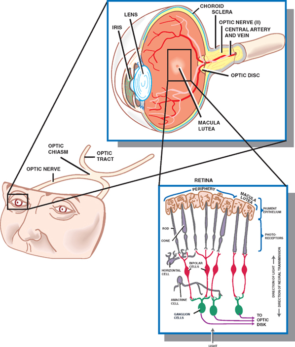
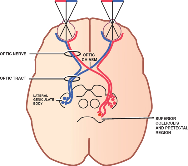
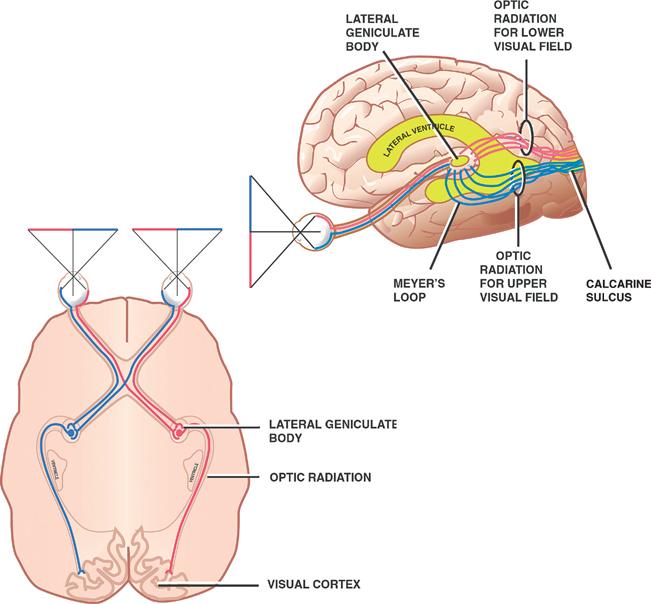
15 Visual System The visual system transforms visual representations of the external world into patterns of neural activity. As a peripheral receptor organ, the eye contains nonneural elements that transmit light stimuli. These stimuli impinge on the neural elements of the eye, which respond by giving rise to impulses that are carried and processed in central pathways. The testing of vision constitutes one of the most sensitive and important parts of the neurologic examination. It is sensitive because the visual pathways traverse the brain’s entire horizontal axis and are thus vulnerable to a wide range of anatomic lesions. It is important because such lesions tend to produce discrete visual deficits that facilitate their localization. See Fig. 15.1. The optic nerve and retina represent outgrowths of the forebrain that are specially adapted for light sensitivity, for modifying light information, and for transmitting the modified information to the thalamus and occipital cortex. The area of the retina where the optic nerve fibers exit the eye is called the optic disc. Because it contains only nerve fibers, the optic disc is a blind spot. Radiating from the center of the disc are the retinal vessels. Roughly two temporal disc diameters away from the optic disc lies the macula lutea, which occupies the center of the retina and is specialized for visual acuity. The greatest visual acuity occurs at a depression in the center of the macula, the fovea centralis, the central part of which contains only cone receptors (one of the two photoreceptor types). The basic cell layers of the retina, from outside in, are the pigment epithelium, rods and cones, bipolar cells, and ganglion cells. Interneurons (i.e., horizontal cells and amacrine cells) and neuroglial cells are also present. There are many more rods than cones. They are more concentrated in the periphery of the retina, and because they exhibit more convergence on bipolar cells (several rods synapsing with a single bipolar cell), they are also more sensitive to low levels of light (e.g., night vision). Cones, which are concentrated more centrally than rods, exhibit less convergence. They sometimes make one-to-one synaptic contacts with bipolar cells, and they therefore provide greater discrimination (i.e., acuity) compared with rods. Cones alone are responsible for color vision. Bipolar neurons connect rods and cones to the ganglion cells. Ganglion cell axons pass dorsally in the optic nerve and terminate in the lateral geniculate body of the thalamus. They transmit information laterally within individual layers of the retina. Fig. 15.1 Retina. See Fig. 15.2. The optic nerve is formed by the myelinated axons of the retinal ganglion cells, which converge on the optic disc and pass through the foramina in the sclera (lamina cribrosa). Although considered a cranial nerve (CN II), the optic nerve is more like a tract within the central nervous system, its myelin sheaths being formed by oligodendrocytes rather than by Schwann cells. Enveloped by the meninges, which fuse with the sclera, the optic nerve leaves the orbit through the optic canal. The central retinal artery and vein are carried in the meningeal sheaths surrounding the optic nerve and are embedded in the ventral portion of the nerve.1 The optic chiasm constitutes a partial crossing of optic nerve fibers. Fibers of the nasal half of each optic nerve decussate to join the uncrossed temporal fibers on the opposite side. The optic tracts, which constitute a continuation of the optic nerves just dorsal to their partial decussation in the optic chiasm, are thus formed by temporal fibers of the ipsilateral optic nerve and nasal fibers of the contralateral optic nerve. In this manner, images in the left field of vision are represented in the right cerebral hemisphere, and vice versa. The optic tract courses dorsolaterally around the cerebral peduncles, where most of its fibers terminate in the lateral geniculate body (LGB) of the thalamus. Other fibers that are specialized for control of eye movements and the pupillary light reflex terminate in the superior colliculus and the pretectal region, respectively. 1Increased intracranial pressure is transmitted to the central vein by its surrounding cerebrospinal fluid. Given a sufficient increase in intracranial pressure, obstruction of the venous return from the retina may develop, thus engorging the optic disc. The funduscopic picture of a disc that is swollen secondary to increased intracranial pressure is referred to as papilledema. Fig. 15.2 Optic nerve, optic chiasm, and optic tract. See Fig. 15.3. The LGB cells that receive impulses from the retinal ganglion cells in turn project axons in the optic radiation (geniculocalcarine tract), which passes dorsally through the retro- and sublenticular parts of the internal capsule and then around the lateral ventricle to terminate in the visual cortex (Brodmann’s area 17) of the occipital lobe. Those fibers of the optic radiation that form the so-called Meyer’s loop pass through the temporal lobe en route to the visual cortex, where they terminate below the calcarine sulcus. The primary visual cortex occupies the medial aspect of the occipital lobe in the region of the calcarine sulcus. As a result of the decussation of nasal fibers at the optic chiasm, each visual cortex receives information only from the opposite visual field. Furthermore, impulses initiated in the inferior retinal quadrants (upper field of vision) are projected on the lower bank of the calcarine sulcus; those initiated in the superior retinal quadrants (lower field of vision) are projected on the upper bank. In sum, cortical representation of the visual field is inverted and reversed from right to left. The primary visual cortex sends impulses to the surrounding association visual cortex (Brodmann’s areas 18 and 19), which is responsible for higher visual functions such as recognition of objects and perception of depth and color.
The Retina
Visual Pathways
Optic Nerve, Optic Chiasm, and Optic Tract
Optic Radiation and Visual Cortex
Related posts:

Stay updated, free articles. Join our Telegram channel

Full access? Get Clinical Tree


