21 Lesions are usually discernible in the mamillary bodies (Fig. 21.1), but may also involve other parts of the hypothalamus, the medial thalamic nuclei, the floor of the third ventricle, the periaqueductal region (Fig. 21.1), the colliculi (Fig. 21.1), the nuclei in the pontomedullary tegmentum (Fig. 21.1) (particularly the dorsal motor nuclei of the vagus), the inferior olives, and the cerebral cortex. Typically, the involved regions are slightly shrunken and show brown discoloration due to hemosiderin deposition, and there may be petechial hemorrhages. The periventricular and periaqueductal lesions often spare a slender strip of subependymal tissue. In some patients, particularly those with previously treated disease, the mamillary bodies may be only mildly discolored and the brain may appear macroscopically normal. 21.1 Wernicke’s encephalopathy. Acute lesions are edematous with relative preservation of neurons, variable necrosis of intervening tissue (Fig. 21.2), and loss of myelinated fibers. Capillaries may appear strikingly prominent due to endothelial hyperplasia and cuffing by macrophages (Fig. 21.2), but this is not a constant feature. There may be petechial hemorrhages and hemosiderin-laden macrophages (Fig. 21.3). Astrocytes show reactive changes. 21.2 Active Wernicke’s encephalopathy. 21.3 Hypothalamic and periaqueductal lesions in acute Wernicke’s encephalopathy. Chronic lesions usually appear gliotic and slightly spongiotic, with mild loss of neurons, depletion of myelinated fibers, and scattered hemosiderin-laden macrophages and astrocytes (Figs 21.4, 21.5). 21.4 Chronic Wernicke’s encephalopathy. 21.5 Chronic Wernicke’s encephalopathy. The histologic changes in Wernicke’s encephalopathy and Wernicke–Korsakoff syndrome are essentially identical. Studies suggest that Korsakoff’s psychosis occurs in those patients who have lesions involving the medial dorsal (or possibly other medial thalamic) nuclei (Fig. 21.6). 21.6 Wernicke–Korsakoff syndrome. The brain and spinal cord appear macroscopically normal. Histologic changes in the CNS occur predominantly in the later stages of pellagra. Betz cells and neurons in the pontine and cerebellar dentate nuclei show striking chromatolysis without associated microglial or astrocytic changes (Fig. 21.7). Other neurons in the brain stem and the anterior horn cells of the spinal cord (Fig. 21.7) may also be affected. Symmetric degeneration of the dorsal columns, especially the gracile funiculi, and, to a lesser extent, of the corticospinal tracts, has been observed. 21.7 Pellagra.
Vitamin deficiencies
THIAMINE DEFICIENCY AND WERNICKE’S ENCEPHALOPATHY
MACROSCOPIC APPEARANCES
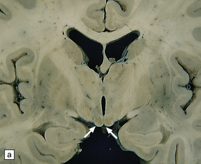
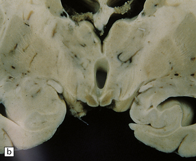
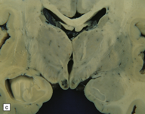
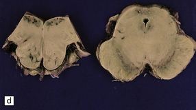
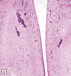
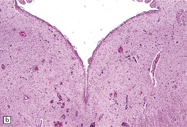
Compare the shape and color of (a) normal mamillary bodies (arrows), with (b) the shrunken, brown mamillary bodies, and (c) the presence of petechial hemorrhages in cases of Wernicke’s encephalopathy. (d) Brown discoloration and petechial hemorrhages in the periaqueductal region, colliculi, and floor of the fourth ventricle.
MICROSCOPIC APPEARANCES
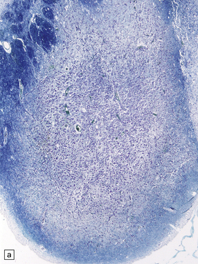
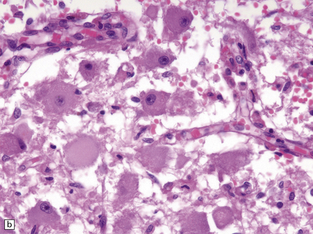
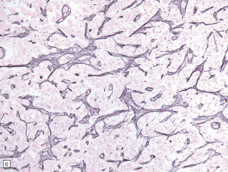
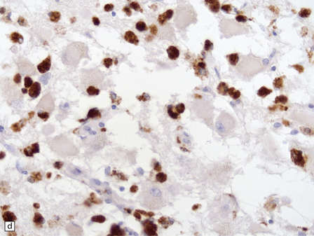
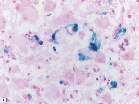
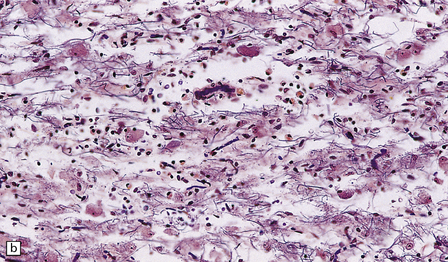
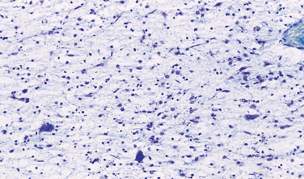
(a) The mamillary body shows loss of myelin and an increase in cellularity. (b) Neuronal somata are relatively preserved, although some neurons are abnormally swollen. (c) The increased prominence of capillaries is well demonstrated by silver impregnation of the capillary basement membranes. (d) The increase in cellularity results mostly from infiltration by macrophages, immunopositive for CD68. (e) Some of the macrophages contain hemosiderin pigment, stained here by Perls’ method.
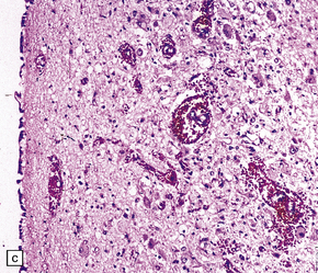
(a) Petechial hemorrhages in the hypothalamus, and (b) periaqueductal gray matter. (c) Higher magnification view of petechial hemorrhages in the hypothalamus.
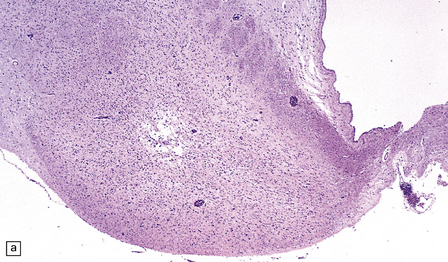
(a) Mild focal spongiosis of the mamillary body. (b) Higher magnification of a gliotic mamillary body.
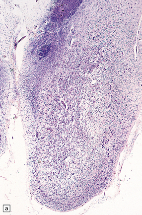
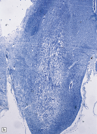
(a) Marked shrinkage and spongy gliosis of the mamillary body. (b) The mamillary body is depleted of myelinated fibers.
Rarefied gliotic medial dorsal thalamic nucleus from a patient with Wernicke–Korsakoff syndrome.
NICOTINIC ACID DEFICIENCY AND PELLAGRA
MACROSCOPIC AND MICROSCOPIC APPEARANCES
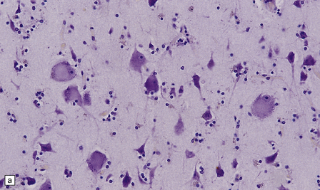
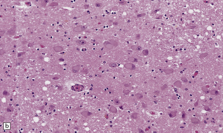
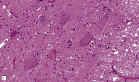
(a) Betz cells in the primary motor cortex are particularly susceptible to chromatolysis in pellagra. (b) Widespread chromatolysis of neurons in the pontine nuclei. (c) Spinal cord in pellagra. Most of the anterior horn cells are chromatolytic.
 NICOTINIC ACID DEFICIENCY AND PELLAGRA
![]()
Stay updated, free articles. Join our Telegram channel

Full access? Get Clinical Tree


Vitamin deficiencies
 WERNICKE’S ENCEPHALOPATHY


 WERNICKE’S ENCEPHALOPATHY


 PELLAGRA


