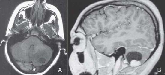
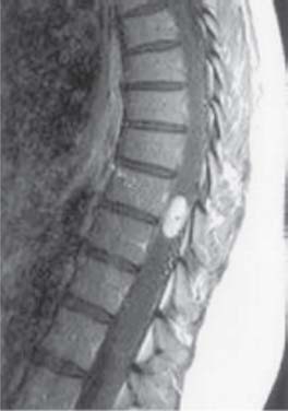
Case 4 Von Hippel–Lindau Disease—Hemangioblastoma Fig. 4.1 (A) T1-weighted magnetic resonance image (MRI) of the brain, axial cut through the posterior fossa. (B) T1-weighted MRI of the brain with contrast enhancement, sagittal cut. Fig. 4.2 T1-weighted magnetic resonance image of the thoracic spine with contrast enhancement, midsagittal cut.
 Clinical Presentation
 Questions
 Answers
< div class='tao-gold-member'>
Only gold members can continue reading. Log In or Register to continue


