6 Olfactory Groove Meningioma

Case 6 Olfactory Groove Meningioma


Stephen J. Hentschel and Lissa Marie Ogieglo


Image

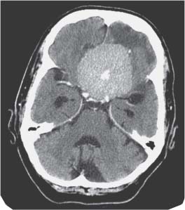
Fig. 6.1 Axial computed tomography (CT) scan of the brain plus contrast.


Image Clinical Presentation


Image Questions


  1. What is your differential diagnosis?
  2. What is your initial management?
  3. What is Foster–Kennedy syndrome?
  4. What further imaging studies, if any, would you request?
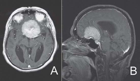
  5. Magnetic resonance imaging (MRI) scan is obtained; please describe the findings (Fig. 6.2).
  6. What are the main features differentiating an olfactory groove meningioma and a tuberculum sellae meningioma?
  7. Describe the pros and cons of the most common potential operative approaches for olfactory groove meningiomas.
  8. Describe the general operative technique including management for the arterial supply and plan for resection.
  9. What is the expected prognosis of this lesion following your treatment? What intraoperative steps can you take to minimize recurrence of the lesion?


Image

Fig. 6.2 (A) Axial T1-weighted magnetic resonance image (MRI) plus gadolinium of the brain. (B) Sagittal T1-weighted MRI plus gadolinium of the brain.


Image Answers


  1. What is your differential diagnosis?

    • Skull-base meningiomas (olfactory groove meningioma, tuberculum sellae meningioma)
    • Pituitary adenoma
    • Craniopharyngioma

  2. What is your initial management?

    • Reversal of warfarin in preparation for operation with vitamin K and fresh frozen plasma
    • Corticosteroid: dexamethasone – loading dose of 10 mg intravenously (i.v.) followed by 4 mg i.v./p.o. (by mouth) every 6 hours
    • Preoperative assessment for fitness of surgery

  3. What is Foster–Kennedy syndrome?

    • Foster–Kennedy syndrome has been described with olfactory groove meningiomas. The components of this syndrome include

      • Anosmia
      • Unilateral optic atrophy
      • Contralateral papilledema

  4. What further imaging studies, if any, would you request?

    • Further imaging studies should include an MRI scan with MR angiography.
    • There is no need for conventional catheter angiography because the relationship of the vessels of the lesion should be well defined in noninvasive angiography.

  5. An MRI scan is obtained; please describe the findings.

    • The T1-weighted MRI sequence with gadolinium demonstrates a large diffusely enhancing tumor arising from the anterior skull base with the anterior cerebral arteries located posterior to the lesion and the optic chiasm located inferior to the lesion.

  6. What are the main features differentiating an olfactory groove meningioma and a tuberculum sellae meningioma?

  7. Describe the pros and cons of the most common potential operative approaches for olfactory groove meningiomas.

    • The most common operative approaches for olfactory groove meningiomas are described below.
    • Subfrontal approach ± orbital osteotomies

      • For larger tumors (> cm), a bicoronal flap is turned.
      • For smaller tumors (< cm), a unicoronal flap is turned.1
      • Advantages

        • Early devascularization along the skull base with division of feeding vessels2
        • Allows for access into orbits to coagulate the ethmoidal arteries that supply the majority of the tumor3
        • Orbital osteotomies minimize frontal lobe retraction.1
        • Allows for harvesting of vascularized pericranium for skull base reconstruction

      • Disadvantages

        • Opens frontal sinus, the increasing risk of postoperative cerebrospinal fluid (CSF) leak and infection2
        • Sacrifice of anterosuperior sagittal sinus

    • Pterional

      • Advantages

        • Early exposure of optic apparatus and carotid artery prior to tumor manipulation1,24
        • Early access to basal cisterns for CSF drainage for brain relaxation4
        • Shorter distance to tumor4
        • Avoids entry into frontal sinus1
        • Spares venous structures2,4
        • Less frontal lobe retraction unless orbital osteotomies are preformed with subfrontal approach1

      • Disadvantages

        • Narrow working angle4
        • May be blinded in upper portion of tumor, which may require extensive frontal lobe retraction4
        • Difficult to access ethmoid arteries2
        • Difficult to repair basal skull defects2

    • Interhemispheric2

      • Advantages

        • Preserves superior sagittal sinus
        • Frontal sinus not opened

      • Disadvantages

        • Higher risk of contusion to frontal lobes
        • Operative route is long and narrow.
        • Risk to bridging veins
        • Difficult to access vascular supply

  8. Describe the general operative technique including management for the arterial supply and plan for resection.

    • Craniotomy ± orbital osteotomies
    • Early interruption of the blood supply

      • If using a subfrontal approach, isolate and cauterize the anterior and posterior ethmoidal arteries within the orbit to reduce the risk of intraoperative hemorrhage.

    • Gentle retraction of the frontal lobes with exposure of tumor1,2
    • The tumor capsule must be dissected, cauterized, and opened. The tumor is then debulked using an ultrasonic aspirator.
    • At the posterior aspect of the tumor, an intact arachnoid plane should be identified separating the tumor from the anterior cerebral arteries, chiasm, and optic nerves.
    • Excellent visualization of the anterior cranial fossa floor to permit tumor resection and repair of defects1

  9. What is the expected prognosis of this lesion following your treatment? What intraoperative steps can you take to minimize recurrence of the lesion?

    • Prognosis

      • Clinically, this patient has a high likelihood of returning to normal mental status with a reversal of her visual changes.1,5
      • It has been shown that these tumors have a high predilection for late recurrence at the cranial base and sinuses with a rate approximated at 30% at 5 years and 41% at 10 years.6
      • Due to the increased difficulty and risks associated with reoperations, reducing the chance of recurrence following the primary resection should be a goal of this surgery4,7
      • Aggressive primary resection including drilling of hyperostotic bone, removal of dura as well as resection of sinus extension is recommended.1 In these circumstances, reconstruction of the skull base is a necessity to prevent postoperative CSF leaks and meningitis.
< div class='tao-gold-member'>

Only gold members can continue reading. Log In or Register to continue

Stay updated, free articles. Join our Telegram channel

Jul 16, 2016 | Posted by in NEUROSURGERY | Comments Off on 6 Olfactory Groove Meningioma

Full access? Get Clinical Tree

Get Clinical Tree app for offline access