7 Sphenoid Wing Meningioma

Case 7 Sphenoid Wing Meningioma


Remi Nader


Image

Fig. 7.1 T1-weighted magnetic resonance imaging sequences with contrast illustrating sphenoid wing meningioma. (A) Axial, (B) sagittal, and (C,D) coronal sections of interest are shown.


Image 
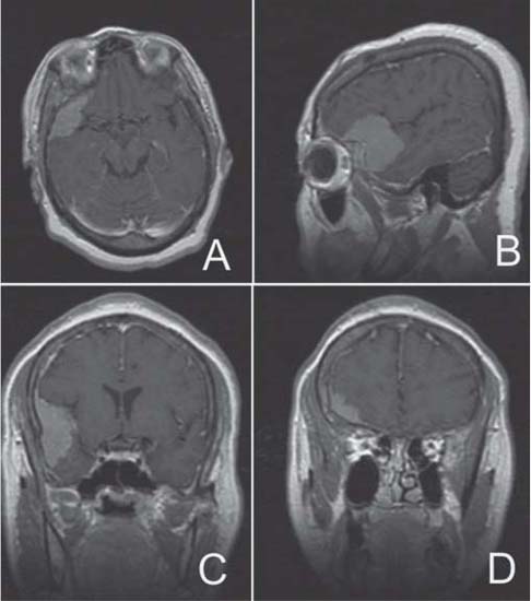
Clinical Presentation


Image Questions


  1. Interpret the MRI scan.
  2. Give a differential diagnosis and the most likely diagnosis.
  3. Give a classification of the most likely diagnosis.
  4. What is your initial management?
  5. How would you approach the lesion surgically?
  6. You decide to resect the lesion via a pterional approach. After the lesion is completely removed, you have a large dural defect. How do you address this defect?
  7. You are able to remove the lesion in its entirety and a surrounding rim of dura up to the level of the sphenoid wing, where you can only cauterize the adherent dural edge, but are unable to resect it. How would you grade your resection and based on what classification?
  8. What are the clinical implications to this grading?
  9. How do you manage her seizures postoperatively?

Image Answers


  1. Interpret the MRI scan.

    • A 4 × 4 × 2-cm right frontotemporal dural-based tumor is seen.
    • The mass is abutting the sylvian fissure.
    • There also appeared to be an attachment along the lateral aspect of the right sphenoid wing.
    • There is some mild mass effect; however, there is no midline shift and no hydrocephalus.

  2. Give a differential diagnosis and the most likely diagnosis.

    • Most likely diagnosis is meningioma.
    • Differential diagnosis

      • Tumor

        • Primary: hemangiopericytoma, primary bony lesion
        • Metastatic lesion

    • Less likely diagnoses include

      • Infection: brain abscess, subdural empyema, meningitis
      • Vascular lesion: arteriovenous malformation
      • Inflammatory condition: sarcoidosis
      • Traumatic: subdural or epidural hematoma

  3. Give a classification based on this location of the most likely diagnosis.

  4. What is your initial management?

    • Admit the patient to the hospital.
    • Obtain preoperative laboratory studies: complete blood count, electrolytes, prothrombin time/partial thromboplastin time, type and cross match 2 to 4 units.
    • Start dexamethasone (6 mg every 6 hours recommended) if vasogenic edema is suspected.
    • Seizure prophylaxis: continue her home regimen or if uncontrolled, change regimen (phenytoin, phenobarbital, or valproate are the recommended options; levetiracetam [Keppra, U.C.B. S.A., Brussels, Belgium] may be used as an adjunct)
    • Obtain preoperative angiogram with possibility of embolization.
    • Obtain medical clearance because of her history of stroke.
    • May also get other imaging studies such as a computed tomography (CT) scan to assess hyperostosis, and magnetic resonance angiography (MRA) if an angiogram is unobtainable.1,2

  5. How would you approach the lesion surgically?

    • Preoperative steps in surgical management

      • Priority is to preserve and improve function.
      • Steroids preoperatively for 48 hours
      • Control blood pressure during anesthesia.
      • Furosemide, mannitol, intravenous antibiotics
      • Head positioning (Mayfield 3-point fixation)

        • Elevate the head above heart level.
        • Head rotation: 30 to 40 degrees
        • Supine, elevated shoulder ipsilateral to tumor

      • Preoperative frameless navigation scan to mark incision and location of the tumor

    • Intraoperative considerations

      • Minimize brain retraction.
      • Plan skin incision to allow full exposure of the tumor.
      • Preserve pericranial tissue attachment to scalp for dural repair.
      • Right pterional or cranio-orbito-zygomatic craniotomy
      • Exposure of lateral sphenoid wing extradurally via bone drilling of the sphenoid
      • Intraoperative ultrasound may be used as an adjunct.
      • Expose as little normal brain as possible.
      • Extensive internal decompression prior to capsule resection
      • Use Cavitron ultrasonic surgical aspirator or cautery loop.
      • Use intraoperative microscope for magnification.
      • Have a self-retaining retractor such as the Budde halo (Integra, Plainsboro, NJ) or Greenberg halo (Codman, Raynham, MA) available.
      • Debulk tumor internally prior to resecting the outer capsule.
      • Remove en block if possible with rim of dura surrounding the tumor.
      • Spare vessels “en passage” especially at the sylvian fissure.
      • Drill hyperostotic bone.

    • Postoperative considerations

  6. You decide to resect the lesion via a pterional approach. After the lesion is completely removed, you have a large dural defect. How do you address this defect?

    • The defect needs to be closed; several options are available.

      • Bovine pericardium
      • Fascia lata (requires preparing a harvesting site)
      • Pericranium (also requires harvesting and potentially expanding the incision)
      • Synthetic dural substitute

    • Closure of the defect may be further optimized by using fibrin glue around the suture line to prevent cerebrospinal fluid seepage.1,2

  7. You are able to remove the lesion in its entirety and a surrounding rim of dura up to the level of the sphenoid wing, where you can only cauterize the adherent dural edge but are unable to resect it. How would you grade your resection and based on what classification?

    • Simpson grade 2 resection
    • Note the Simpson grading scale for meningioma resection5,6:

      • Grade 1 – Resection complete with the dural attachment
      • Grade 2 – Coagulation of dural attachment
      • Grade 3 – Resection of tumor only with dural attachment left behind
      • Grade 4 – Subtotal resection
      • Grade 5 – Decompression

  8. What are the clinical implications to this grading?

    • There is a 20% rate of recurrence at 5 years with Simpson grade 2 resections.5,6

  9. How do you manage her seizures postoperatively?

    • Keep her on her preoperative antiepileptic regimen if this was controlling seizures before the surgery.
    • Do not discontinue for at least 6 months to 1 year until you are sure she is seizure free.
    • Frequent follow-ups (monthly) to check antiepileptic levels
    • May elect to get the neurology service involved
    • Seizure control can be achieved in up to 88% of patients undergoing complete resection of meningioma.7
< div class='tao-gold-member'>

Only gold members can continue reading. Log In or Register to continue

Stay updated, free articles. Join our Telegram channel

Jul 16, 2016 | Posted by in NEUROSURGERY | Comments Off on 7 Sphenoid Wing Meningioma

Full access? Get Clinical Tree

Get Clinical Tree app for offline access