Acquired Peripheral Neuropathies


Acquired Peripheral Neuropathies


Thomas H. Brannagan III

Kurenai Tanji


APPROACH TO PERIPHERAL NERVE DISORDERS

The peripheral nervous system is composed of multiple cell types and elements that subserve diverse motor, sensory, and autonomic functions. The clinical manifestations of neuropathies depend on the severity, distribution, and functions affected. Peripheral neuropathy and polyneuropathy are terms that describe syndromes resulting from diffuse lesions of peripheral nerves, usually manifested by weakness, sensory loss, pain, and autonomic dysfunction. Mononeuropathy indicates a disorder of a single nerve often resulting from local trauma, compression, or entrapment. Mononeuropathy multiplex signifies focal involvement of two or more nerves, usually as a result of a generalized disorder such as diabetes mellitus or vasculitis. This chapter discusses an approach to peripheral nerve disorders, mononeuropathies, including plexus disorders, and specific acquired polyneuropathies. Inherited neuropathies are discussed in Chapter 88.


EPIDEMIOLOGY

Peripheral neuropathy which includes polyneuropathies of various causes and mononeuropathies are common disorders. One mononeuropathy, carpal tunnel syndrome, is estimated to occur in 3% to 5.8% and is three times more common in women than men. A prevalence of 2% to 7% of symmetric polyneuropathy has been found in studies conducted around the world. Currently, the most common cause of peripheral neuropathy worldwide is diabetes, although prior to 1994, the most common cause of peripheral neuropathy worldwide was leprosy. The prevalence of peripheral neuropathy increases with age, affecting 15% of the population older than the age of 40 years and 24% older than the age of 70 years.


CLINICAL FEATURES

Polyneuropathy may occur at any age, although particular syndromes are more likely to occur in certain age groups. Charcot-Marie-Tooth (CMT) disease, for example, often begins in childhood or adolescence, whereas neuropathy associated with paraproteinemia is seen more frequently with increasing age. The onset and progression differ; the Guillain-Barré syndrome (GBS), tick paralysis, and porphyria begin acutely and may remit. Others, such as vitamin B12 deficiency or carcinomatous neuropathy, begin insidiously and progress slowly. Still others, such as chronic inflammatory demyelinating polyneuropathy, may begin acutely or insidiously and then progress with remissions and relapses.

The myelin sheaths or the motor or sensory axons or neurons themselves may be predominantly affected, or the neuropathy may be mixed, axonal, or demyelinating. Most polyneuropathies, especially those with primary demyelination, affect both motor and sensory functions. A predominantly motor polyneuropathy is seen in lead toxicity, dapsone or n-hexane intoxication, tick paralysis, porphyria, some cases of GBS, and multifocal motor neuropathy. Sensory neuropathy is divided into loss of large-diameter and small-diameter nerve fibers, although a combination of fiber types is most typical. Predominantly small-fiber neuropathy, often with concomitant autonomic dysfunction, is seen in diabetes mellitus, amyloidosis, Fabry disease, and lepromatous leprosy. Less prevalent, predominant large-fiber neuropathy occurs with thallium poisoning, paraneoplastic ganglioneuritis, Sjögren disease, pyridoxine (vitamin B6) toxicity, and syphilis. Predominant involvement of the autonomic system can be seen in acute or chronic autonomic neuropathy or in amyloidosis.

Symptoms of polyneuropathy include distal pain, paresthesias, weakness, and sensory loss. Pain may be spontaneous or elicited by stimulation of the skin and may be sharp or burning. Paresthesias are usually described as numbness (a dead sensation), tingling, buzzing, stinging, burning, or a feeling of constriction. Lack of pain perception may result in repeated traumatic injuries with degeneration of joints (arthropathy or Charcot joints) and in chronic ulcerations.

Weakness is greatest in distal limb muscles in most neuropathies; there may be paralysis of the intrinsic foot and hand muscles with footdrop or wrist-drop. Tendon reflexes are often lost, especially in demyelinating neuropathy. In severe polyneuropathy, the patient may become quadriplegic and respirator dependent. The cranial nerves may be affected, particularly in GBS and diphtheritic neuropathy. Cutaneous sensory loss appears in a stockingand-glove distribution. All modes of sensation may be affected, or there may be selective impairment of “large” myelinated fiber functions (position and vibratory sense) or “small” unmyelinated fiber functions (pain and temperature perception). Often, detection of painful stimuli is impaired with a delayed and greater than normal reaction. Inappropriate pain perception is often paradoxically present despite loss of pain fibers.

Involvement of autonomic nerves may cause miosis (small pupil), Adie pupils, anhidrosis (impaired sweating), orthostatic hypotension, sphincter disturbance, gastrointestinal dysmotility, impotence, and vasomotor abnormalities; these may occur without other evidence of neuropathy but are more commonly seen in association with symmetric distal polyneuropathy. Diabetes mellitus is the most common cause. Amyloidosis causes notably severe autonomic neuropathy. The majority of neuropathy types produce distal impairment of sweating, vasomotor reflexes, and local influences that help produce the typical trophic signs in the feet of neuropathy patients. Distal loss of sweating may induce symptomatic excessive sweating proximally as a compensatory response.

In mononeuropathy or mononeuropathy multiplex, focal motor, sensory, and reflex changes are restricted to areas innervated by specific nerves. When multiple distal nerves are affected in mononeuropathy multiplex, the pattern may coalesce into more symmetric involvement suggesting polyneuropathy. The most frequent causes of mononeuropathy multiplex are vasculitic neuropathy, diabetes mellitus, rheumatoid arthritis, brachial neuropathy,
leprosy, or sarcoidosis. Asymmetric neuropathy is also seen in multifocal motor neuropathy with conduction block, sometimes with increased anti-GM1 antibody titers, multifocal demyelinating sensory and motor neuropathy (Lewis-Sumner syndrome), and brachial neuritis.

Processes that affect nerve roots are termed radiculopathies and affect a single or multiple myotomes or dermatomes. Focal compression is most common, but other processes also target nerves at these proximal sites. Electric-like pain radiating down a specific segment is most characteristic; loss of sensation or strength in a specific territory occurs with more severe involvement. Plexopathy occurs with compromise of the brachial or lumbosacral plexus and often displays a pattern of multiple contiguous nerves or nerve root impairment.

Superficial cutaneous nerves may be thickened and visibly enlarged secondary to Schwann cell proliferation and collagen deposition from repeated episodes of segmental demyelination and remyelination or from amyloid or polysaccharide deposition in the nerves. Hypertrophic nerves may be observed or palpated in the demyelinating form of CMT disease (type I), Dejerine-Sottas neuropathy, Refsum disease, von Recklinghausen disease (neurofibromatosis), and various other disorders.

Fasciculations, or spontaneous contractions of individual motor units, are visible twitches of limb or cranial muscles. They are characteristic of anterior horn cell diseases but also occasionally occur in other chronic neuropathic conditions. Fibrillation potentials are discharges of denervated single muscle fibers and are not visible on the skin but are recordable during electromyography (EMG). Myokymia is worm-like muscle activity seen in limb muscles in a small number of disorders including radiation plexopathy, episodic ataxia type 1, and certain autoimmune channelopathies. Potassium channel antibodies or defects are implicated in some entities; facial myokymia is more common and less specific.


MONONEUROPATHIES AND COMPRESSION NEUROPATHIES


THE SPINAL ROOTS AND BRACHIAL PLEXUS

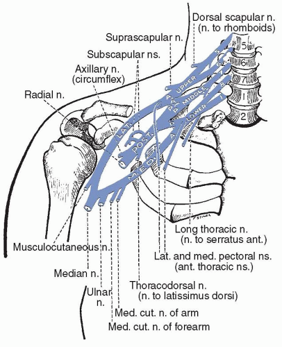
At each cervical spinal level, numerous rootlets containing both motor and sensory fibers join after leaving the spinal cord to form the spinal roots that exit the spinal canal through the intervertebral foramen of the spinal column, immediately branching into anterior and posterior rami. The nerve roots are commonly injured by degenerative joint disease and disk herniation at the cervical and lumbosacral levels. Importantly, the dorsal root ganglia (i.e., the cell bodies of the sensory nerves) are located outside the foramen and are spared in foraminal compression, meaning the remainder of the sensory nerve will remain viable and will appear normal on nerve conduction studies, even though it is disconnected from the central nervous system (CNS) and the patient reports numbness and pain. Before forming the brachial plexus, the C5 nerve root gives off a proximal branch, the dorsal scapular nerve (to the rhomboid muscles), whereas the C5, C6, and C7 roots give proximal branches that join to form the long thoracic nerve, supplying the serratus anterior muscle. The C5 and C6 roots then join to form the upper trunk of the brachial plexus, whereas the C7 root forms the middle trunk, and the C8 and T1

roots form the lower trunk. The upper trunk gives off a small branch, the suprascapular nerve, which supplies the supra- and infraspinatus muscles. All trunks pass through the supraclavicular fossa under the cervical and scalene muscles. Each trunk then forms two branches and these branches regroup to form new divisions, the cords, as they course through the thoracic outlet, between the first rib and the clavicle, along with the subclavian artery. The lateral branches of the upper and middle trunks contribute to the lateral cord (i.e., C5, C6, C7), whereas the medial branches join with the lateral branch of the lower trunk and move dorsally to form the posterior cord (i.e., C5, C6, C7, C8). Finally, the lower trunk gives rise to the medial cord (i.e., C8, T1). The lateral and medial pectoral nerves branch off near the juncture of the trunks and the lateral and medial cords, respectively, supplying the pectoralis major muscle.


TABLE 87.1 Neuropathy Diagnosis and Laboratory Tests


Cause or Diagnosis


Manifestations


Laboratory Tests


Vitamin deficiency/excess


S, SM, SYM


Vitamins B12, pyridoxine (B6), B1, folate, vitamin E, methylmalonic acid


Infectious


Lyme disease


S, SM, SYM, MF, CN


Serology, PCR


HIV-1


S, SM, SYM, MF, CN


Serology, PCR


Hepatitis C


S, SM, SYM, MF, CN


Serology, PCR


Herpes zoster


S, radicular


Serology, PCR


CMV


SM, M, SYM, MF


Serology, PCR, culture


Immune mediated


Guillain-Barré and variants


SM, S, M, SYM, MF, CN


IgG antiganglioside antibodies (GM1, GD1a, GQ1b, GD1b), urine porphyrins


IgM antibody associated


M, MF


IgM anti-GM1, GD1a


S, SM, SYM


IgM anti-MAG, sulfatide, GD1b, GQ1b


Monoclonal gammopathy


M, S, SM, SYM, MF


Serum immunofixation electrophoresis, quantitative immunoglobulins


Autonomic neuropathy


Autonomic dysfunction


Antinicotinic acetylcholine receptor antibodies, anti-Hu


Vasculitis


SM, S, MF, SYM


ESR, cryoglobulins, hepatitis C serology, or PCR


Sarcoidosis


SM, S, MF, SYM


ACE, chest radiograph


Celiac disease


S, SM, MF, SYM


Antigliadin, endomysial, transglutaminase antibodies


Rheumatologic diseases


SM, S, MF, SYM


SSA-Ro, SSB-La antibodies


Sjögren syndrome


ANA, ANCA (PR3, myeloperoxidase), dsDNA, Ab, RNP, rheumatoid factor


Lupus


Wegener granulomatosis Rheumatoid arthritis


Paraneoplastic


Lung cancer


S, SYM


Anti-Hu Ab, chest radiograph/CT


Waldenström syndrome


SM, S, M, SYM, MF


Serum immunofixation electrophoresis


Myeloma


SM, M, SYM, MF


Serum and urine immunofixation electrophoresis, skeletal survey


Hereditary


CMT-1


Demyelinating, SM, SYM, MF


DNA tests for PMP-22, MPZ, EGR2, Cx32, others


CMT-2


Axonal, SM, SYM


DNA tests for NF-L, Cx32, MPZ, others


Mitochondrial


NARP, SM, MF


Serum lactate, thymidine phosphorylase, DNA testing


Other


Axonal, S, SM, amyloid, porphyria


DNA tests for transthyretin, periaxin, urine porphyrins


Metabolic/toxic


Diabetes


S, SM, SYM, MF, CN


Fasting glucose, HgbA1c, glucose tolerance test


Renal failure


S, SM, SYM


Chem 7


Thyroid disease


S, SM, SYM, MF


TSH, T4


Heavy metal toxicity


S, SM, SYM, MF


Urine lead, mercury, arsenic, thallium


S, sensory; SM, sensorimotor; SYM, symmetric; MF, multifocal; CN, cranial nerves; PCR, polymerase chain reaction; CMV, cytomegalovirus; GM1, GD1a, ganglioside components of myelin; M, motor; MAG, myelin-associated glycoprotein; ESR, erythrocyte sedimentation rate; ACE, angiotensin-converting enzyme; SSA, SSB, antigens for Sjögren syndrome severe antibodies; ANA, antinuclear antibody; ANCA, antineutrophil cytoplasmic antibodies; RNP, ribonucleoprotein; CT, computed tomography; PMP, peripheral myelin protein; MPZ, myelin protein zero; NARP, neuropathy, ataxia, and retinitis pigmentosa; EGR, early growth response protein; Cx32, connexin; NF-L, neurofilament light chain; HgbA1c, hemoglobin A1c; TSH, thyroid-stimulating hormone.


TABLE 87.2 American Academy of Neurology Recommendations for Testing for Distal Symmetric Polyneuropathy


Basic Laboratory Evaluation for Distal Symmetric Neuropathy


CBC, ESR, or CRP


Vitamin B12a and if B12 is low normal, metabolites, including methylmalonic acid and/or homocysteine.a


Comprehensive metabolic panel, including fasting blood glucosea, glucose tolerance testa if indicated to look for impaired glucose tolerance


Serum protein immunofixation electrophoresisa


Urine analysis, urine electrophoresis


Inquire about drugs and toxins


Other Laboratory Testing that May Be Performed in Selected Patients


ANA, rheumatoid factor, anti-Ro/SSA, anti-LA/SSB, antineutrophil cytoplasmic antigen (ANCA) antibodies, cryoglobulins


Campylobacter jejuni, cytomegalovirus (CMV), hepatitis panel (B and C), HIV, Lyme antibodies, herpesviruses tests, West Nile virus tests, cerebrospinal fluid (CSF) analysis


Antigliadin; IgA transglutaminase antibodies; endomysial antibodies; vitamins E, B1, and B6


Serum angiotensin-converting enzyme (ACE), CSF analysis with CSF ACE


Arsenic, lead, mercury, thallium


Antiganglioside antibodies (GM1, GD1a, GD1b, GD3, GQ1b, GT1b), anti-MAG, paraneoplastic antibodies (anti-Hu, anti-CV2), CSF oligoclonal bands


Molecular tests for Charcot-Marie-Tooth, Hereditary neuropathy with tendency to pressure palsy (HNPP), familial amyloidosis


Skeletal survey; computed tomography or MRI of chest, abdomen, or pelvis; ultrasound of abdomen and pelvis; positron emission tomography (PET), CSF cytology


a Tests with highest yield.


CBC, complete blood count; ESR, erythrocyte sedimentation rate; CRP, C-reactive protein; IgA, immunoglobulin A; MAG, myelin-associated glycoprotein; MRI, magnetic resonance imaging.


Data from England JD, Gronseth GS, Franklin G, et al. Practice parameter: evaluation of distal symmetric polyneuropathy: role of laboratory and genetic testing (an evidence-based review). Neurology. 2009;72:185-192.


The thoracodorsal nerve that supplies the latissimus dorsi and the subscapular nerve that supplies the teres major each branch medially off the posterior cord. The posterior cord persists distally, becoming the radial nerve, after giving off a smaller lateral branch, the axillary nerve, which supplies the deltoid. The lateral and medial cords then each contribute a branch to form the median nerve, composed of the medial branch of the lateral cord and the lateral branch of the medial cord, joined in the middle of the plexus. The lateral branch of the lateral cord persists distally, becoming the musculocutaneous nerve, whereas the medial branch of the medial cord becomes the ulnar nerve (Fig. 87.1; Tables 87.3 and 87.4).

The brachial plexus may be injured by traumatic, neoplastic, infectious, radiation, and other processes. Careful history and neurologic examination, in concert with a detailed understanding of plexus anatomy, is the first step in recognizing plexus injury and differentiating it from injury to the nerve roots or peripheral nerves. Electrophysiologic assessment with EMG and nerve conductions is often critical in confirming the diagnosis, and imaging studies may also be indicated. Mixed syndromes of radicular plexus and peripheral nerve injury may also occur, making localization even more challenging. The roots or trunks of the brachial plexus may be damaged by lacerations, gunshot wounds, or direct trauma. They may be compressed by tumors or aneurysms or stretched and torn by violent movements of the shoulder in falls, dislocation of the shoulder, carrying heavy loads on or over the shoulder, and by traction during birth. The syndromes of the roots and trunks cause deficits principally in the distribution of the affected nerve roots. Partial paralysis and incomplete sensory loss are common because many muscles of the arm receive innervation from two or more roots. Compression at the level of the thoracic outlet (thoracic outlet syndrome) is addressed separately in Chapter 78.


FIGURE 87.1 The brachial plexus. (From Haymaker W, Woodhall B. Peripheral Nerve Injuries. Philadelphia: WB Saunders; 1945.)


TABLE 87.3 Innervation of the Muscles of the Shoulder Girdle


Muscle


Nerve


Spinal Nerve Roots


Sternocleidomastoid


Accessory


X1, C2, C3


Trapezius


Accessory


C3, C4


Serratus anterior


Long thoracic


C5, C7


Levator scapulae


Dorsal scapular


C5, C6


Rhomboideus major


Dorsal scapular


C5, C6


Rhomboideus minor


Dorsal scapular


C5, C6


Subclavius


Subclavian


C5, C6


Supraspinatus


Suprascapular


C5, C6


Infraspinatus


Suprascapular


C5, C6


Pectoralis major


Medial and lateral pectoralis


C5, C6


Pectoralis minor


Medial pectoralis


C5, C6


Teres major


Subscapular


C5, C6


Latissimus dorsi


Thoracodorsal


C6, C7


Subscapularis


Subscapular


C5, C6


Deltoid


Axillaris


C5, C6


Teres minor


Axillaris


C5, C6


Trunk and Root Injury


UPPER RADICULAR SYNDROME

Upper radicular syndrome (Erb or Erb-Duchenne palsy) results from damage to the upper roots (C4, C5, or C6) or the upper trunk. Such lesions are most commonly the result of stretch injuries during difficult deliveries, especially when forceps are used, and cause paralysis of the deltoid, biceps, brachioradialis, pectoralis major, supraspinatus, infraspinatus, subscapularis, and teres major muscles in varying combinations. If the lesion is near the roots, the serratus anterior, rhomboids, and levator scapulae are also paralyzed. Clinically, this causes weakness of flexion at the elbow and of abduction and internal and external rotation of the arm. There is also weakness or paralysis of apposition of the scapula and backward-inward movements of the arm. Sensory loss is incomplete and consists of hypesthesia on the outer surface of the arm and forearm. The biceps reflex is absent. Unless treated by passive range-of-motion exercise, these patients may develop chronic contractures with the arm extended at the side, fully adducted, and pronated, with the hand flexed and facing rearward (e.g., the waiter’s tip position).


MIDDLE RADICULAR SYNDROME

Middle radicular syndrome results from damage to the seventh cervical root (C7) or the middle trunk. Such lesions cause paralysis primarily of the muscles supplied by the radial nerve except the brachioradialis, which is entirely spared. Clinical weakness parallels that of injury to the radial nerve below the level of its branch to the brachioradialis. Sensory loss is variable and when present is limited to hypesthesia over the dorsal surface of the forearm and the external part of the dorsal surface of the hand.


TABLE 87.4 Innervation of Muscles of the Arm and Forearm


Muscle


Nerve


Root


Biceps brachii Brachialis


Musculocutaneous


C5, C6


Triceps


Radialis


C7, C8


Anconeus


Radialis


C7, C8


Brachioradialis


Radialis


C5, C6


Extensor carpi radialis


Radialis


C6, C7


Pronator teres


Medianus


C6, C7


Flexor carpi radialis


Medianus


C7, C8


Palmaris longus


Medianus


C7, C8


Flexor digitorum sublimis


Medianus


C7, C8


Flexor digitorum profundus


Medianus, ulnaris


C7, C8


Flexor carpi ulnaris


Ulnaris


C7, C8


Supinator


Radialis


C7, C8


Extensor digitorum communis


Radialis


C7, C8


Extensor digiti minimi


Radialis


C7, C8


Extensor carpi ulnaris


Radialis


C7, C8


Abductor pollicis longus and brevis


Radialis


C7, C8


Extensor indicis proprius


Radialis


C7, C8


LOWER RADICULAR SYNDROME (KLUMPKE PALSY)

Lower radicular syndrome (Klumpke palsy) results from injury to the lower trunk or lower roots (C7-T1), which causes paralysis of the flexor carpi ulnaris, the flexor digitorum, the interossei, and the thenar and hypothenar muscles. This pattern mimics a combined lesion of the median and ulnar nerves. Clinically, a flattened or simian hand is seen, with loss of all intrinsic hand musculature and with loss of sensation on the inner side of the arm and forearm and on the ulnar side of the hand. The triceps reflex is lost. If the communicating branch to the inferior cervical ganglion is injured, there is paralysis of the sympathetic nerves, causing a Horner syndrome.


Cord Injury

Lesions of the cords cause motor and sensory loss resembling that seen after injury to two or more peripheral nerves. Lateral cord injury causes weakness in the distribution of the musculocutaneous nerve and the lateral head of the median nerve, including weakness in the pronator teres, flexor carpi radialis, and flexor pollicis. Posterior cord injury causes weakness paralleling that resulting from combined damage to the radial and axillary nerves, whereas medial cord injury mimics combined damage to the ulnar nerve and the medial head of the median nerve (finger-flexion weakness).


Diffuse Plexus Injury

Generalized injury to the brachial plexus is usually unilateral but occasionally appears bilaterally. Such injury results from a more diffuse polyneuropathy, such as chronic inflammatory demyelinating
neuropathy, or from multifocal motor neuropathy. A variety of insults may produce injury selectively affecting the brachial plexus, but tumor infiltration, radiation plexitis, and idiopathic plexitis are among the most important. Almost any neoplasm with a propensity for the chest may affect the plexus, but those cancers originating locally, such as lung and breast cancer, are most likely to cause injury. Such tumors may cause extrinsic compression of the plexus as they grow or may directly infiltrate the nervous tissue. Other neoplasms, such as lymphoma, may infiltrate the plexus and cause progressive deficits without any apparent mass effect or enlargement of the plexus itself in the initial stages. Magnetic resonance imaging (MRI) with contrast is the best way to confirm these lesions.

Idiopathic brachial plexitis (also known as the Parsonage-Turner syndrome or neuralgic amyotrophy) usually begins with a sudden, sharp pain affecting one shoulder, often with radiation down the ipsilateral arm, later followed by arm or shoulder weakness. The pain persists for hours or a few days with gradual improvement and usually resolves completely within days to weeks, leaving some sensory and motor dysfunction. It may be bilateral or asymmetric. Localization is often difficult, because plexus involvement ranges from diffuse to multifocal, and often includes patchy injury to the nerve branches off the plexus (e.g., the axillary nerve).

Electrodiagnostic studies, if performed at least 14 to 21 days after onset, usually localize the injury to the plexus but may demonstrate multifocal involvement. Patterns of both axonal and demyelination injury have been reported. The diversity of physiologic disorders in different nerves or even within the same nerve is attributed to involvement of the terminal nerve twigs or to patchy damage of discrete bundles of fibers within the cords or trunks of the brachial plexus or its branches. The long thoracic and anterior interosseous nerves are commonly affected. Autoimmune or infectious causes have been suggested, but the etiology is obscure. Some cases have occurred in small epidemics, and the disorder may follow intravenous heroin use, HIV seroconversion, surgery, and delivery.

There is no clear evidence that immunosuppressive therapy alters the course of the disease. However, short courses of tapering oral steroids are often prescribed if the patient presents shortly after symptom onset. Variants of this syndrome have also been described, including one with isolated, pure sensory injury affecting the lateral, antebrachial, cutaneous, and the median nerves.

A hereditary form that is frequently recurrent and bilateral is rarely encountered. Lumbosacral plexitis also occurs but much less frequently. Recovery depends on the severity of the initial insult. Although most patients recover well over 6 to 12 months, some are left with permanent disability. It is considered good in about 66%, fair in 20%, and poor in 14%. Clinical recovery may take 2 months to 3 years.


Thoracic Outlet Syndrome

The term thoracic outlet syndrome (TOS) encompasses different syndromes that arise from compression of the nerves in the brachial plexus or blood vessels (i.e., subclavian or axillary arteries, or veins in the same area). The putative compression sources are also diverse. How often these lesions are actually responsible for symptoms and how the symptoms should be treated are matters of intense debate. Studies done mainly by orthopedists, vascular surgeons, and neurosurgeons have included reports on several hundred patients who were treated surgically for this syndrome. When neurologists write about the neurogenic form of TOS, however, the tone is always skeptical, and the syndrome is described as exceedingly rare, with an annual incidence of about 1 per 1 million persons.


PATHOLOGY

The T1 and C8 nerve roots and the lower trunk of the brachial plexus are exposed to compression and angulation by anatomic anomalies that include cervical ribs and fibrous bands—of uncertain origin. Other nearby structures, such as scalene muscles, are dubious sources of compression. Cervical ribs are commonly found in asymptomatic people, and it is therefore difficult to assume that the presence of a cervical rib necessarily explains local symptoms. In addition to the neural syndromes, the same anomalies may compress local blood vessels and cause vascular syndromes, usually separated into arterial and venous entities. These conditions are also rare and may cause neurogenic symptoms by distal nerve ischemia but not pressure on the brachial plexus. Some cases follow local trauma.


CLINICAL FEATURES

Patients have pain in the shoulders, arms, and hands, or sometimes in all three locations. Hand pain is often most severe in the fourth and fifth fingers. The pain is aggravated by use of the arm and arm “fatigue” may be prominent, meaning local discomfort after brief effort. There may or may not be hypesthesia in the affected area.

Critics have divided cases into two groups: the true neurogenic TOS and the disputed syndrome. In the true syndrome, there are definite clinical and electrodiagnostic abnormalities. This disorder is rare and is almost always caused by a taut fibrous band extending from a cervical rib or abnormally elongated C7 transverse process; the band stretches the distal C8 and T1 roots or lower brachial plexus trunk. There is unequivocal wasting and weakness of hand muscles innervated by these segments. Changes are almost always unilateral.

EMG and nerve conduction studies demonstrate a pattern of low-amplitude median motor, ulnar sensory, and medial antebrachial cutaneous-evoked responses. Ulnar motor responses to hypothenar muscles may be involved to a lesser degree. EMG signs of active and chronic denervation are limited to involved muscles, most severely in the abductor pollicis brevis, and are attributed to a major contribution from T1.

In the disputed form, there are no objective motor or sensory signs or consistent laboratory abnormalities. Attempts to reproduce the syndrome by passive abduction of the arm (i.e., the Adson test) or other maneuvers have been cited, but the same abnormalities may be found in normal people and have no specific diagnostic value. The diagnosis is usually made by the treating surgeon; symptoms are frequently bilateral and complicated by legal or other nonmedical issues.

Similarly, studies of the application of electrodiagnostic techniques have not been blinded or controlled, so less specific abnormalities have been noted; the findings include isolated abnormalities of ulnar sensory nerve amplitude, conduction velocity after stimulation of the Erb point, ulnar F waves, and ulnar somatosensory-evoked potentials. MRI may show deviation or distortion of nerves or blood vessels, bands extending from the C7 transverse process, or other local anomalies. MRI quantitative estimates of the size of the thoracic outlet may show smaller than average dimensions, as were the cases in blinded reviews of series that compared vascular and neurogenic patients together with controls but do not prove that the differences are causative. Magnetic resonance angiography and Doppler ultrasonography may help assess possible vascular compression.


THE SPINAL ROOTS AND LUMBAR AND SACRAL PLEXI


The Lumbar Plexus

The spinal roots at L2, L3, and L4 join to form the lumbar plexus in the psoas major muscle. This plexus gives off a number of principally sensory nerves, including the iliohypogastric, the ilioinguinal, the genitofemoral, and the lateral femoral cutaneous. The femoral nerve is derived from the L2, L3, and L4 roots, passing to the anterior leg along the lateral aspect of the psoas muscle (which it supplies); exiting the pelvis; and passing under the inguinal ligament to supply the pectineus, sartorius, rectus femoris, vastus lateralis, vastus intermedius, and vastus medialis muscles; and terminates as the pure sensory saphenous nerve in the medial lower leg.

The obturator nerve arises from anterior branches of the L2, L3, and L4 roots, forming in the psoas muscle and entering the pelvis anteriorly to the sacroiliac joint. It passes through the obturator canal, branching anteriorly to supply the adductor longus and brevis, and the gracilis, as well as posteriorly to supply the obturator externus, and half of the adductor magnus. It carries sensation from a small and variable area on the inner surface of the medial thigh, knee, and occasionally just below the medial knee.


The Sacral Plexus

The sacral plexus is formed from the L5, S1, and S2 roots, with variable contributions from L4. The superior gluteal nerve arises from the L4, L5, and S1 roots and supplies the gluteus medius and minimus and tensor fascia lata; the inferior gluteal nerve arises from the L5 and S1 roots and supplies the gluteus maximus.

The sciatic nerve is formed from the posterior fusion of the L4, L5, and S1 roots, exiting the pelvis via the greater sciatic foramen and passes through or under the piriformis muscle. It is functionally divided into a lateral fibular portion, which supplies the short head of the biceps femoris, and a medial tibial portion, which supplies the long head of the biceps femoris, the semitendinosus, and the semimembranosus. The nerve divides into the common fibular (formerly called the peroneal) and tibial nerves above the posterior knee.

The common fibular nerve branches laterally from the sciatic trunk in the popliteal fossa then moves superficially to wind around the head of the fibula. It then divides into the superficial fibular nerve to supply the peroneus longus and peroneus brevis and the deep fibular nerve, which supplies the tibialis anterior, extensor hallucis longus, peroneus tertius, and extensor digitorum brevis. The tibial nerve supplies the gastrocnemius and soleus, tibialis posterior, flexor digitorum longus, and flexor hallucis longus. It descends through the lower leg between the medial malleolus and the flexor retinaculum, dividing into the medial plantar nerve to supply the abductor hallucis, flexor digitorum brevis and flexor hallucis brevis, and the lateral plantar nerve to supply the abductor digiti minimi, flexor digiti minimi, abductor hallucis, and interosseous muscles. Both the tibial and fibular nerves supply sensory branches, which join to form the sural nerve below the popliteal space.


Radiation Plexopathy

Irradiation for carcinoma may damage nervous tissue, especially with high-voltage therapy. Brachial plexopathy is seen after radiotherapy for breast cancer; caudal roots and lumbosacral plexus are sometimes affected by radiation therapy for testicular cancer or Hodgkin disease. The first symptom is usually severe pain, followed by paresthesia and sensory loss. There may be a latent period of 12 to 20 months; in milder cases, several years may elapse before symptoms appear. Limb weakness peaks many months later. Latency intervals of up to 20 years have been reported. The damage may affect a single peripheral nerve initially and then progress slowly to involve others. Clinically, tendon reflexes disappear before weakness and atrophy becomes obvious; fasciculation and myokymia may be prominent. EMG and conduction studies reveal evidence of axonal damage; myokymic discharges are characteristic and can help differentiate plexopathy caused by radiation from plexopathy caused by tumor infiltration. High-resolution MRI is also potentially useful. No effective treatment is known. Radiation-induced fibrosis and microvascular injury are suspected mechanisms.


THE PROXIMAL NERVES OF THE ARM


The Axillary Nerve

The axillary nerve is the last branch of the posterior cord of the brachial plexus before forming the radial nerve. It arises from C5 and C6, supplies the deltoid and teres minor muscles, and transmits cutaneous sensation from a small patch on the lateral shoulder. Axillary neuropathy may be caused by trauma, fracture, humeral head dislocation, and brachial plexitis. Weakness of arm abduction after the first 15 to 30 degrees of movement is typical. Outward, backward, and forward movements of the arm are also weakened, although less dramatically. Sensory loss is limited to a small patch over the lateral deltoid.


The Long Thoracic Nerve

The long thoracic nerve arises from C5, C6, and C7 and supplies the serratus anterior muscle. This nerve is most commonly injured in isolation by forceful, downward pressure on the shoulder, which
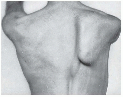
stretches and compresses it. Typically, such pressure is caused by carrying excessively heavy loads on the shoulder (e.g., furniture, carpets, heavy sacks, backpacks slung over one shoulder, etc.), although it may also appear after acute impact, such as that occurring while playing football. A more archaic term, although one still in use, is hod-carrier’s palsy, in reference to the hod or container bricklayers formerly placed on the shoulder to carry bricks up to roof tops when constructing chimneys. Injury of this nerve destabilizes the scapula, causing winging, and prevents the rotation of the scapula needed to enable the last few degrees of abduction of the arm from 90 to 180 degrees over the head. Injury following acute or chronic trauma is characterized by weakness in elevation of the arm above the horizontal plane. Winging of the scapula is most prominent when the arm is fully abducted or elevated anteriorly (Fig. 87.2). Winging is often not readily apparent with the arm resting at the side.


FIGURE 87.2 Paralysis of the serratus anterior muscle with winging of the scapula.


The Brachial Cutaneous and Antebrachial Cutaneous Nerves

The brachial and antebrachial cutaneous nerves branch directly from the C8 to T1 plexus and provide sensation to the medial arm and upper two-thirds of the forearm. These nerves are usually injured in conjunction with the medial cord of the brachial plexus and are rarely injured in isolation.


The Suprascapular Nerve

The suprascapular nerve fibers arise from C5 and C6, ultimately branching from the upper trunk of the brachial plexus. The primarily motor nerve innervates the supraspinatus and infraspinatus muscles. Affected patients have difficulty moving the arm from the side through the first 15 to 30 degrees of abduction and with external shoulder rotation. Shoulder trauma or more diffuse brachial plexus injury is most common; isolated nerve injury is rare.


THE PERIPHERAL NERVES


The Radial Nerve

The radial nerve is a continuation of the posterior cord and contains elements of the C5, C6, C7, C8, and T1 nerve roots. It is predominantly a motor nerve and innervates the chief extensors of the forearm, wrist, and fingers. It descends through the axilla to supply the triceps, giving off three minor sensory branches to the upper arm, then winds posteriorly around the humerus in the spiral groove. After exiting the spiral groove, the nerve innervates the brachioradialis and extensor carpi radialis longus muscles then moves laterally to enter the forearm between the brachialis and brachioradialis muscles. There, it branches into a primary sensory component, the superficial radial nerve, which supplies sensation to the dorsoradial aspect of the distal forearm and the dorsal surface of the hand, and a motor component, the posterior interosseous nerve, which supplies all the remaining forearm extensor muscles and often the supinator as well (Table 87.5).

The clinical findings after radial nerve injury depend on the level of the lesion. Injury in the axilla, classically caused by improperly fitting crutches that are too long, causes triceps weakness as well as weakness of the remaining radial myotome and numbness in the radial dermatome. Injury in the spiral groove caused by humeral fracture or extrinsic compression (e.g., Saturday night palsy) causes weakness of the radial myotome below the elbow, with prominent wrist-drop, weakness of finger extension, and sensory loss in the distribution of the superficial radial nerve but preserved elbow extension. Mild elbow flexor weakness may be present as a result of involvement of the brachioradialis, which should be easy to distinguish on physical examination. The posterior interosseous branch may also be injured by entrapment as it passes through the supinator muscle in the tight space of the arcade of Frohse.

Injury of the posterior interosseous nerve spares the brachioradialis and the extensor carpi radialis longus, as well as the superficial radial nerve, causing radial deviation of the wrist with attempted wrist extension but no sensory loss (i.e., posterior interosseous neuropathy). Damage to the superficial radial branch may occur at the wrist as a result of tight-fitting jewelry or handcuffs, causing pure sensory loss over the dorsum of the hand without weakness. Evaluation of radial nerve injury often includes electrodiagnostic studies and may include imaging studies, depending on the site of the lesion. Treatment focuses on relieving the cause of compressive injury, if possible. Posterior interosseous nerve syndrome is sometimes treated with surgical release.


The Median Nerve

The median nerve derives from the C6 through T1 nerve roots, passing through the lateral and medial cords of the brachial plexus, which each contributes a segment to the nerve. The median nerve passes down the arm and through the two heads of the pronator teres at the level of the forearm, ultimately supplying the pronator teres as well as the flexor carpi radialis, palmaris longus, and flexor digitorum superficialis muscles. It then branches into the pure
motor anterior interosseous nerve, which supplies the flexor pollicis longus, pronator quadratus, and flexor digitorum profundus I and II, and into a main branch, which passes through the carpal tunnel, further branching into the recurrent thenar nerve, supplying the abductor and the lateral flexor pollicis brevi and the opponens pollicis before terminating in the palm, where it supplies lumbricals I and II. Pronation is mediated by the pronator quadratus and pronator teres, wrist flexion by the flexor carpi radialis and palmaris longus, flexion of the thumb and the index and middle fingers by the superficial and deep flexors, and opposition of the thumb by the opponens pollicis (Table 87.6).


TABLE 87.5 Muscles Innervated by the Radial Nerve


Triceps


Extensor digiti minimi


Anconeus


Extensor carpi ulnaris


Brachioradialis


Abductor pollicis longus


Extensor carpi radialis longus and brevis


Extensor pollicis longus and brevis


Supinator


Extensor indicis proprius


Extensor digitorum communis


TABLE 87.6 Muscles Innervated by the Median Nerve


Pronator teres


Pronator quadratus


Flexor carpi radialis


Abductor pollicis brevis


Palmaris longus


Opponens pollicis


Flexor digitorum sublimis


Flexor pollicis brevis


Flexor digitorum profundus


Lumbricales (digits one and two)


Flexor pollicis longus


The median nerve supplies sensation to the radial side of the palm, the ventral thumb, index and middle fingers, the radial half of the ring finger, as well as the dorsal surfaces of the distal phalanx of the thumb, and the middle and terminal phalanges of the index and middle fingers. Isolated lesions of the median nerve cause weakness and sensory loss in the aforementioned distributions, but only a few movements are paralyzed because of the synergistic contributions of muscles innervated by other nerves to these movements. However, there may be absence of flexion in the index finger and near-complete paralysis of the opponens pollicis. The median nerve may be injured by trauma, ischemia, and other processes but most commonly is damaged by anatomic compression. It may be entrapped between the heads of the pronator teres muscle, causing weakness and sensory loss in the above distributions, with sparing of the pronator teres itself, which is innervated more proximally (i.e., the pronator teres syndrome).

Only gold members can continue reading. Log In or Register to continue

Stay updated, free articles. Join our Telegram channel

Jul 27, 2016 | Posted by in NEUROLOGY | Comments Off on Acquired Peripheral Neuropathies

Full access? Get Clinical Tree

Get Clinical Tree app for offline access