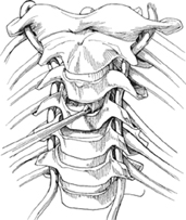
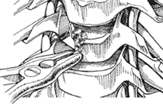
18 James C. Farmer Anterior cervical corpectomy is a method of decompressing the cervical spinal cord and nerve roots. It is performed by removing the disk above and below the vertebral body that is to be resected, followed by the vertebral body itself (complete corpectomy) (Fig. 18.1). Under some circumstances only one disk is removed along with a portion of the adjacent vertebral body/bodies (partial corpectomy). Fig. 18.1 Anterior diskectomy. Anterior osteophytes, ossification of the posterior longitudinal ligament, as well as disk herniations positioned behind the vertebral body can be resected utilizing careful surgical technique. Excellent decompression of the spinal cord and nerve roots can be achieved. Sagittal deformities such as kyphosis can be corrected through this technique. Following a corpectomy, all anterior compression of the spinal cord and nerve roots should be relieved. If kyphosis exists preoperatively, it can be corrected allowing restoration of normal cervical lordosis. On occasion, supplemental posterior stabilization may be required. Magnetic resonance imaging (MRI) remains the modality of choice when treating patients with cervical disorders. MRI with flexion and extension sagittal images can be helpful in evaluating positional effects on cervical stenosis as well as evaluating the reduction of instability. Computed tomography (CT), which can be combined with myelography, provides the best imaging of bony structures. A CT scan is paramount when evaluating ossification of the posterior longitudinal ligament (OPLL) to adequately assess the degree of ossification as well as the number of levels involved. Myelography can evaluate the effects of flexion and extension dynamically on spinal cord and nerve root compression. The patient is positioned supine with the arms at the side and carefully padded and protected. A roll is placed between the shoulders to allow the head to be positioned in a neutral to a slightly extended position. It is important to avoid overextension of the neck as this can cause spinal cord impingement and possible injury. Gardner-Wells tong traction can be used. Spinal cord monitoring is recommended. In patients with significant spinal cord compression or instability, fiberoptic intubation may be necessary. Obtaining proper imaging studies as well as a careful preoperative evaluation of these studies is paramount for optimal surgical treatment. Determination of the number of levels involved as well as the extent of compression at each individual level is essential. Additionally, careful evaluation of the location and course of the vertebral artery is necessary to avoid iatrogenic injury. At the time of surgery, complete diskectomies prior to resection of the vertebral bodies facilitate assessment of the depth of the vertebral body as well as the location of the spinal canal. In cases of ossification of the posterior longitudinal ligament where the ossification is extremely adherent to the dura, direct resection can be dangerous. Successful decompression can be performed by removing the posterior longitudinal ligament on either side of the ossified area, and allowing it to float away anteriorly from the cord (anterior floating technique) without necessitating direct resection, and high risk of dural tear and subsequent spinal fluid leak. When performing the corpectomy, a high-speed burr can be used to resect most of the vertebral body, leaving only a thin rim of posterior cortical bone. The posterior cortical bone can be removed using either a small curette or a Kerrison rongeur. It is imperative to identify the uncovertebral joints bilaterally when performing the decompression. This enables the surgeon to maintain orientation to the midline, facilitating an adequate decompression of the spinal canal, and avoiding dissecting too laterally with the risk of injury to the vertebral artery. Careful resection of the posterior cortex and posterior longitudinal ligament is necessary to avoid injury to the dural sac and spinal cord. Significant bleeding can occur during resection of the vertebral body and can make visualization difficult at times. This can be controlled with the judicious use of bone wax and hemostatic agents such as Gelfoam, Surgicel, and Avitene. During the performance of multilevel corpectomies, adequate surgical exposure is paramount. During surgical exposure, the raising of subplatysmal flaps as well as the takedown of fascial structures along the surgical interval provide optimal exposure and enable mobilization of the soft tissue structures along the anterior spine. The operating room microscope provides optimal illumination and excellent visualization of the surgical field for both the surgeon and the assistant. Loupe magnification and a headlight also enable safe neural decompression. Standard diskectomies should be performed above and below the vertebral body to be resected. Visualization of the posterior longitudinal ligament and uncovertebral joints on each side is necessary for an adequate decompression. The initial portions of the corpectomy can then be performed either with a rongeur or with various high-speed burrs (Fig. 18.2). The width of the decompression should be on average 15 to 16 mm. Again, identification of the uncovertebral joints during the decompression facilitates assessment of the widest extent of decompression that can be performed should it be needed. The posterior longitudinal ligament does not routinely need to be excised unless it is felt that a compressive lesion posterior to the posterior longitudinal ligament exists or in the case of an OPLL. With OPLL the surgeon may elect to remove the entire ossified ligament, or in cases where it is felt to be exceedingly adherent to the dura, the anterior floating method can be utilized (Fig. 18.3).
Anterior Cervical Corpectomy
Description
Key Principles
Expectations
Indications
Contraindications
Special Considerations
Special Instructions, Position, and Anesthesia
Tips, Pearls, and Lessons Learned
Difficulties Encountered
Key Procedural Steps
Related posts:

Stay updated, free articles. Join our Telegram channel

Full access? Get Clinical Tree


