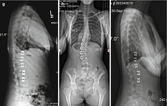
Fig. 43.1
Demonstration of spinal mobility after T10–L3 VBS
43.1.1 Historical Overview
Stapling across physes of the long bones has been accepted as an effective method for treating limb malalignment in young children for over 50 years [20, 21]. Around the same time, the potential benefits were discovered for the spine. Animal studies using a rat tail model confirmed the ability to modulate vertebral growth plates with skeletal fixation devices [22]. In 1951, Nachlas and Borden [23] were initially optimistic about their ability to create and correct lumbar scoliosis in a canine model using a staple that spanned several vertebral levels. Many of the dogs exhibited some correction, and some of the animals exhibited arrest of their curve progression. Some of the staples failed because they spanned three vertebrae. The enthusiasm for this new treatment was lost after the application of their stapling technique in three children with progressive scoliosis that yielded poor results. Other investigators have, similarly, been dissatisfied with convex stapling as a means of controlling progressive scoliosis.
Results for humans with congenital scoliosis were presented as early as 1954 [24], but the results were disappointing. The scoliosis correction was limited because the children had little remaining growth, and the curves were severe, with considerable rotational deformity. Some staples broke or loosened, possibly because of motion through the intervertebral disks. While the concept of stapling the anterior vertebral end plates/physes for growth modulation and curve stabilization seemed sound, the staples designed for epiphyseal stapling about the knee were prone to dislodging in the spine because they were not designed to function across the intervertebral disk and accommodate to the movement of the functional spinal unit.
In 2003, our institution published the results of a patient cohort that had undergone anterior VBS for moderate adolescent idiopathic scoliosis (AIS) with a newly designed staple [25]. The significance of the study was demonstrated by 87 % of the curves that were maintained using the stapling technique.
43.1.2 Basic Science Overview
Recent work has shown the efficacy of anterior growth modulation in animals. Despite the successful use of staples for epiphysiodesis of long bones in angular deformity, staples for growth modulation around the spine were not nearly as successful. The obvious issue was that staples designed for the long bones were prone to dislodge in the spine because they were not designed for movement which occurs in the spine.
Medtronic Sofamor Danek (Memphis, Tennessee) designed staples using nitinol, a shape memory alloy, which have 510(k) approval from the FDA specifically for fixation in the anterior spine within a single vertebral body or for fixation of hand and foot osteotomies. These staples are unique in that the prongs are straight when cooled but clamp down into the bone in a “C” shape when the staple returns to body temperature, thus providing secure fixation. The nitinol staple described in this text is considered “off-label” by the FDA. Nitinol is a biocompatible shape memory metal alloy composed of 50 % nickel and 50 % titanium. The temperature at which the staples will undergo the shape transformation can be controlled by the manufacturing process. Injury to surrounding tissues through the transformation temperature has not been seen in animal or human experience with cervical spinal fusions.
Nitinol has a very low corrosion rate and has been used in orthodontic appliances. Implant studies in animals have shown minimal elevations of nickel in the tissues in contact with the metal; the levels of titanium are comparable to the lowest levels found in tissues of titanium hip prostheses, and titanium is considered a biologically safe implant material. No method of sterilization used in operating rooms has been shown to have any effect on the metal’s properties. Although sensitivity to nickel occurs in a very low percentage of the population, it is not anticipated to occur through the use of the nitinol staple. The crystal structure in nitinol is different than the small amount of nickel crystal structure in stainless steel such that the nickel does not leach out in nitinol compounds as it can on occasion with stainless steel. The nitinol staple has been tested in a goat scoliosis model applied across a disk space by Braun et al. [26] and has been shown to be safe and have utility for arresting iatrogenic curves of less than 70° in the goat.
43.1.3 Clinical Outcomes
In 2003, Betz and colleagues [25] reported on the use of the nitinol staples in 21 skeletally immature patients with AIS. Indications for the procedure were either brace noncompliance, often due to psychosocial reasons, or curve progression despite bracing. They found the procedure to be safe and effective, with the results comparable to the expected results of bracing. In 2005, this same group [27] reported on 39 patients and their increased experience with the procedure. Stabilization of the curve was seen in 87 % of those patients older than 8 years at the time of stapling who had a curve of 50° or less with at least 1 year of follow-up. No curve less than 30° at the time of stapling progressed more than 10° at follow-up.
In 2010, Cuddihy et al. reported a retrospective study comparing VBS to bracing for patients with moderate idiopathic scoliosis using identical inclusion criteria [28]. In this comparison of two cohorts of patients with high-risk (Risser 0–1) moderate idiopathic scoliosis (measuring 25–44°), the results of treatment of smaller thoracic curves (25–34°) by VBS were statistically better than the results seen with bracing (82 % versus 54 %, respectively, p = 0.05) when the cohorts were adjusted for mean age (10.5 years). For thoracic curves measuring 35–44°, the results were poor in both groups. The results of lumbar VBS and bracing were similar for curves measuring 25–44°. This study suggests that VBS can be used as an alternative or adjunct to bracing for patients with certain curve sizes who are noncompliant with bracing (Figs. 43.2 and 43.3).


Fig. 43.2
PA (a) and lateral standing radiographs (b) of a 12-year-old female demonstrating a 21° thoracic curve and a 38° thoracolumbar curve. (c) Bone age shows the patient to be Sanders 3. Preoperative bending radiographs (d, e) demonstrate the flexibility of the curve. Patient underwent right-sided thoracoscopic VBS from T10 to L3. Her first standing radiographs (f, g) demonstrated thoracic curve correction to 10° and lumbar curve correction to 9°. Latest follow-up (h, i) at 3 years post-op demonstrates a thoracic curve of 14°, lumbar curve of 18° degrees, and improvement of thoracic kyphosis to 27°


Fig. 43.3
AP (a) and lateral standing radiographs (b) of an 8-year-old female demonstrating a 34° thoracic curve. Preoperative standing radiograph (c) demonstrates the flexibility of the curve. Patient underwent right-sided thoracoscopic VBS from T6 to L1. Her first standing radiographs (d, e) demonstrated thoracic curve correction to 20°. Latest follow-up (f, g) at 2.5 years post-op demonstrates a thoracic curve of 15° and improvement of thoracic kyphosis to 36°
Another study with 2-year outcome data consisted of 41 curves (26 thoracic, 15 lumbar) [29]. Thirteen patients had both curves stapled. The mean age was 9.4 years. Curves decreasing by greater than 10° were considered “improved.” Curves within 10° of their preoperative measurement were considered “no change,” and those progressing greater than 10° were considered “worse.” Success was defined as “improved” or “no change.” Thoracic curves measuring less than 35° had a 79 % success rate. Curves measuring less than 20° on first standing radiograph had an 86 % success rate. In patients with thoracic curves greater than 35°, 6 of 8 progressed past 50°. Seventy-one percent of patients with hypokyphosis showed improvement to a normal sagittal profile. One patient demonstrated worsening of kyphosis associated with coronal progression. Lumbar curves had an overall 87 % success rate, with only one patient with a preoperative curve of 40° progressing to 50°. Five patients lost greater than 10° of lordosis, but the final lumbar lordosis remained in the normal range. Complications were minimal, with a mean blood loss of 214 cc. A recent study by Auriemma et al. (unpublished data) reviewed the results of 63 patients who underwent VBS between the ages of 7 and 15 years for moderate idiopathic scoliosis (average Cobb, 30°). Of the 24 patients (36 curves) who reached skeletal maturity, defined as Risser grade 4 or 5, 71 % (12/17) of thoracic and 89 % (17/19) of lumbar curves were treated “successfully,” defined as either greater than 10° improvement in Cobb angle or within 10° of preoperative curve magnitude.
VBS has shown superior outcomes when curves correct to less than 20° on first standing radiographs [29, 30]. Subanalyses of lumbar curves from these studies revealed higher success rates in curves that corrected to less than 20° on first standing radiograph (88.9 %) than those that did not (83.3 %) [29, 30]. As a result of these findings, the treatment algorithm for VBS has been transitioning from stabilization of preoperative curves to maintenance of intraoperative correction by the addition of postoperative nighttime bracing. Recently, a cohort of AIS patients with moderate (20–45°) thoracolumbar/ lumbar (TL/L) curves underwent VBS and adjuvant postoperative bracing (unpublished data). TL/L Cobb angle significantly improved from a mean of 34° preoperatively to 21° at a minimum of 2-year follow-up. Lateral trunk shift also improved from a mean of 1.8 cm preoperatively to 0.5 cm at most recent follow-up. Although health-related quality of life outcomes have not correlated with trunk shift [31], trunk imbalance has been shown to negatively impact self-image and is a major clinical concern of patients.
Based on this review, we have altered our strategy for when to use staples alone and when to use additional strategies, as follows: if the thoracic curve measures 35–45° and does not bend below 20°, then we will offer vertebral body tethering or a posterior hybrid distraction implant, a unilateral VEPTR, or a growing rod in addition to stapling (Fig. 43.4). If on the first standing radiograph the curve does not measure below 20°, we will brace the child until the curve measures less than 20°.


Fig. 43.4
PA (a) and lateral standing (b) radiographs of a 13-year-old female who underwent T7-T11 VBS and hybrid rod placement (c, d)
43.2 Clinical and Technical Overview
43.2.1 Indications and Contraindications
Patients who have at least 1 year of growth remaining, a scoliosis deformity for which brace treatment would be considered, or who may have failed or refused bracing, are good candidates for the stapling procedure. Lenke 1, 3, 5, and 6 scoliosis curves are ideal for treatment with vertebral stapling. Other indications are as follows: age less than 13 years for girls and less than 15 for boys; Risser 0 or 1, at least 1 year of growth remaining by wrist x-ray, or Sanders digital stage less than or equal to 4; thoracic curves 25–35°, and lumbar coronal curves less than or equal to 45°, with minimal rotation and flexible to less than or equal to 20°; and sagittal thoracic curve less than or equal to 40°.
Medical contraindications are the same as for any anterior spine or chest procedure and include systemic infection, active respiratory disease such as uncontrolled asthma, or conditions with increased anesthetic risk. Significantly compromised pulmonary function may be a relative contraindication.
We do not perform vertebral stapling for lumbar curves over 45° or for thoracic curves over 35° that do not bend to less than 20° because our early experience has yielded poor results. For these larger curves we now perform vertebral body tethering as described recently by Samdani et al. [32]. Over 100 vertebral body tethering procedures have been performed at our institution with 12 for primary lumbar curves. Initial results demonstrate 50 % initial correction with gradual improvement as the child grows. Also, if the curve on the first erect film does not measure less than 20°, the patient should wear a corrective nighttime brace until it does. Kyphosis greater than 40° is also a relative contraindication because of the potential for the creation of hyperkyphosis with growth. We occasionally do a lateral Stagnara view to confirm true kyphosis if there is a question.
Surgeons with experience in anterior spine surgery and especially minimally invasive techniques should be able to perform this procedure. It may be helpful to enlist the assistance of an experienced general or thoracic surgeon. With the use of thoracoscopic and minimally invasive techniques for lumbar curves, scoliotic vertebrae from T3 to L4 can be stapled while limiting the total scar length. Placement of instrumentation at other levels will depend on anatomic variances in the location of the subclavian, azygous, or iliac vessels and the size of the psoas muscle. As a general rule, we try to avoid stapling the L3–L4 disk because of the risk to the nerve roots if a transpsoas approach is used or if retracting the psoas to get the staple posterior to midline of the body requires significant psoas mobilization and vessel ligation.
43.2.2 Technical Overview
43.2.2.1 Equipment/Instrumentation
Bipolar cautery, monopolar cautery.
Nitinol staples: staples are straight when cooled and clamp down into the bone in a C shape when achieving body temperature.
Thoracoscope.
Basin with sterile ice water.
Fluoroscopy.
43.2.2.2 Anesthesia and Positioning
General anesthesia and intubation with a double-lumen endotracheal tube for thoracic curves
Lateral decubitus position with convex side of the scoliosis in the “up” position
Soft pads under all pressure points
C-arm under table for PA and lateral imaging.
All vertebral bodies included in the Cobb angle of the curve are instrumented. Under single-lung general anesthesia, patients are placed in the lateral decubitus position with the convex side of the scoliosis curve in the “up” position. The table is not flexed, and only a small axillary roll is placed. Patient positioning is critical and can be used to maximize correction. The axillary role is often positioned at the apex of the proximal thoracic curve, several centimeters lower than in the standard lateral decubitus position, in order to allow the main thoracic curve to partially correct.
This procedure lends itself to the use of minimally invasive surgical techniques. If video-assisted thoracoscopy is being utilized for insertion, then one-lung ventilation will be necessary, unless carbon dioxide (CO2) gas insufflation is available to displace the lung for visualization of the spine and surrounding structures. Using fluoroscopy, a lateral image of the patient can be utilized to confirm the levels of the vertebra and also to center the ports over the midportion of the vertebral bodies (Fig. 43.5). The standing position is based on the surgeon’s preference, but in our practice the surgeon usually stands in front of the patient with the access surgeon or the assistant holding a camera next to the surgeon. A second assistant stands on the opposite side to help with the retraction (Fig. 43.6).


Fig. 43.5
(a) The patient is placed in a lateral decubitus position. (b, c) Fluoroscopy is used to confirm the levels of the vertebrae and to center the ports over the midportion of the vertebral bodies

Fig. 43.6
Surgeon positioning during VBS. The surgeon usually stands anterior to the patient with the access surgeon or the assistant holding a camera next to the surgeon. A second assistant stands on the opposite side to help with the retraction
In the thoracic spine, most incisions will be close to or within the area of the posterior axillary line. The first port is made in the fifth to seventh intercostal space along the anterior axillary line for visualization with the scope. Additional ports, generally 2 or 3, are made in the posterior axillary line for insertion of the staples. Two oblique incisions are usually required for placement of six or more staples (Fig. 43.7). The incisions are about 2.5–3 cm long and are oblique in such a way as to follow the slope of the ribs. Each incision can then be used to make two to three internal intercostal ports. This allows several levels to be stapled through each skin incision and accommodates the size of the instruments and implants. Fluoroscopy or direct visualization with the thoracoscope are both reliable methods for planning the incision.


Fig. 43.7
Generally, 2 but up to 4 ports in the posterolateral line are used, with the thoracoscope being inserted in the anterior axillary line at the apex of the curve. Incisions are oblique and follow the slope of the ribs
Staples that cross the thoracolumbar junction require partial reflection of the diaphragm anteriorly. Lumbar disk spaces can usually be exposed with a retroperitoneal mini-open approach through a single incision. The incision length is 2.5–3 cm and, similar to the thoracic spine, is localized based on the image intensifier. During the approach, the psoas is either retracted posteriorly or carefully separated longitudinally directly over the posterior half of the disk under EMG control [33]. We generally place the staples posterior to the midline of the lumbar vertebral bodies, ligating or mobilizing the segmental vessels and retracting the psoas. A posterior staple will theoretically avoid diminishing lordosis of the lumbar spine. Position of the staples is reconfirmed using fluoroscopy at the end of the procedure.
While the patient is in the lateral decubitus position, often the flexible main thoracic curve reduces. To further reduce the curve while placing the staples, lateral pressure can be applied through an inserter affixed to staples previously placed in another level. This trial inserter can be used to push the spine straight, thus maximizing correction on the operating table. This may be important because patients who have less than or equal to 20° of curvature on the first erect radiographs typically have better maintenance of correction.
Stay updated, free articles. Join our Telegram channel

Full access? Get Clinical Tree


