, Peter O. Newton2, 3 , Christine L. Farnsworth2 and Kevin Parvaresh3
(1)
Department of Orthopaedics, University of Utah, Salt Lake City, UT, USA
(2)
Division of Orthopedics, Rady Children’s Hospital, San Diego, San Diego, CA, USA
(3)
Department of Orthopaedic Surgery, University of California, San Diego, San Diego, CA, USA
Keywords
Growing spine imagingEOS imaging systemRadiation risk in childrenKey Points
Innovative imaging technology, such as the EOS system, allows for postural assessment of spinal morphology.
Advances in three-dimensional (3D) imaging are being utilized to improve deformity progression analysis, surgical planning, and translational research potential.
Functional imaging such as dynamic MRI shows promise for a novel method for assessment of outcomes in the management of pediatric deformity.
New technology for 3D analysis is significantly reducing radiation exposure in children.
9.1 Introduction
Management of deformity and disease in a child with a growing spine presents unique challenges to the treating physician. Since the introduction of X-rays by Wilhelm Roentgen in 1895, there has been a steady improvement in our ability to quantify and analyze spinal deformity. More advanced imaging techniques now make it possible to understand these deformities in three dimensions, allowing for a better understanding of strategies for three-dimensional (3D) correction.
There have been significant advances in our ability to image spine and chest deformity in growing children. Rather than simply measuring the nature and degree of deformity, imaging can be used to assess volume and function in children who are unable to cooperate with normal test such as pulmonary function studies. This is being done with dynamic magnetic resonance imaging (MRI) and a significant reduction of exposure using the EOS system. The purpose of this chapter is to provide an overview of current imaging techniques for managing children with deformity in the growing spine.
9.2 Plain Radiography
The initial evaluation of suspected spinal deformity in a child begins with a thorough history and physical examination. If there is clearly a spinal deformity present, then plain radiographs should be obtained. It is the responsibility of the requesting physician to specifically tell the imaging department the type of film needed. When possible, these are best taken as orthogonal views in the upright or standing position (posterior–anterior (PA) and lateral) (Fig. 9.1). The PA exposure lessens the amount of radiation exposure to the breast tissue and reproductive organs. The entire spine from C1 to the pelvis should be included as a single image and should include the entire chest wall. Many spinal deformities in children impact the shape and volume of the chest. Rib anomalies and fusions are easily seen, and they provide a sense of relative chest hypoplasia. Many congenital deformities in the thoracic spine shorten the spine and result in constricted lung volumes. Hypoplasia of the chest restricts normal alveolar multiplication during growth and may result in thoracic insufficiency syndrome as described by Campbell et al. [1].
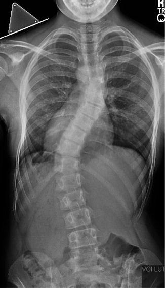
Fig. 9.1
PA radiograph of the spine. The PA radiograph allows for assessment of diagnosis, curve type, magnitude, coronal balance, and skeletal maturity. The PA view has significantly less radiation exposure to sensitive tissues including the thyroid and breasts
Plain radiographs allow for assessment of the etiology of the deformity (idiopathic, congenital, neuromuscular, etc.) and analysis of both coronal and sagittal balance. Curve severity can be measured and documented by measuring the various curves using the Cobb technique. Skeletal maturity is assessed by looking at various growth plates such as the tri-radiate cartilage of the hip or the iliac apophysis (Risser Sign). It is valuable to have a visible marker on the film to account for magnification and allow for accurate measurements of spinal height and growth over time. This allows for measurement of spinal growth using T1-S1 heights. Campbell et al. [1] described the space available for the lung (SAL), a ratio of the distance from the top of the lung to the apex of the diaphragm, comparing left to right (Fig. 9.2a, b). This measurement may have direct implications on the development of the lung.
Fig. 9.2
(a) Space available for the lung (SAL). This is a ratio that documents the effect of the three-dimensional deformity of the chest on potential lung growth. (b) Space available for the lung (SAL). The SAL is a ratio expressed as a percentage of the distance from the diaphragm to the apex of the lung (A/B) measured on an upright radiograph of the chest when comparing one side to the other. In this example, the SAL is 70 % when comparing the right to the left
Differences in the relative angle of the ribs are referred to as rib-vertebral angle differences (RVADs). An RVAD >20° in children with idiopathic early onset scoliosis is felt to correlate with a greater risk of the curve becoming progressive during growth rather than spontaneous correction that may be seen when the RVAD is <20°. An RVAD >20° might warrant an earlier use of correction techniques such as elongation-derotation casting.
9.2.1 EOS System
The EOS system (EOS Imaging, Paris, France) is a recent advancement in imaging technology that allows three-dimensional (3D) assessment of scoliosis deformity through the acquisition of two-dimensional (2D) images. The system utilizes two orthogonal X-ray beams moving in unison with particle detectors along the length of the imaging area, producing two linked orthogonal radiographs. Three-dimensional reconstruction is then performed using SterEOS® software (EOS Imaging, Paris, France), in which a 3D base template is manipulated to produce an anatomic 3D model (Fig. 9.3). The base template for this technology was generated using contour detection from 2D radiographs and subsequently validated with CT images using a synthetic spine phantom model [2]. The template is contoured to match multiple discrete points measured manually on the 2D radiographs to generate the final 3D model (Fig. 9.4). The resultant 3D model may then be manipulated in all three planes based on the operator’s preference for assessment.
Fig. 9.3
 Anesthesia and Postoperative Management of Spinal Deformity Surgery in Growing Children
Regulatory Policies Regarding Pediatric Spinal Devices
Convex Growth Arrest for Congenital Scoliosis
Management of Spine Tumors in Young Children
Growth Modulation Techniques: Titanium Clip-Screw Implant System (HemiBridge)
Single and Dual Traditional Growing Rods
Anesthesia and Postoperative Management of Spinal Deformity Surgery in Growing Children
Regulatory Policies Regarding Pediatric Spinal Devices
Convex Growth Arrest for Congenital Scoliosis
Management of Spine Tumors in Young Children
Growth Modulation Techniques: Titanium Clip-Screw Implant System (HemiBridge)
Single and Dual Traditional Growing Rods


Biplanar imaging (EOS Imaging, Paris, France) of a 4-year 4-month-old child with early onset scoliosis. (a) Vertebrae are identified by the operator. The spine is then detected by the software (SterEOS®). Next, a template is superimposed, which the operator adjusts to match the radiograph contour. (b) Radio view, (c) contours view, and (d) surface views of the adjusted template are shown
Related posts:
Anesthesia and Postoperative Management of Spinal Deformity Surgery in Growing Children
Regulatory Policies Regarding Pediatric Spinal Devices
Convex Growth Arrest for Congenital Scoliosis
Management of Spine Tumors in Young Children
Growth Modulation Techniques: Titanium Clip-Screw Implant System (HemiBridge)
Single and Dual Traditional Growing Rods
Stay updated, free articles. Join our Telegram channel
Full access? Get Clinical Tree
