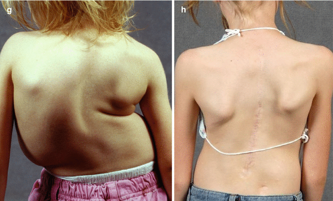
Fig. 36.1
Patient is a 2 + 9-year-old female with infantile idiopathic scoliosis. (a, b) She presented with a 126° scoliosis and thoracic kyphosis deformity. She had a normal total spine MRI and a negative genetics evaluation. She was placed in preoperative halo-gravity traction and underwent a five-level anterior apical release and fusion. (c, d) She then had further traction reducing her scoliosis to 54°. (e, f) Her 5.5-year postoperative x-rays show good maintenance of deformity correction. (g, h) Her preoperative and postoperative clinical photographs demonstrate her much improved appearance
As the chronologic age of the patient increases, the indications for spinal fusion with progressive deformity increase as well. Although an EOS patient becomes an adolescent at 10 years of age, performing a spinal fusion for progressive scoliosis in a 9-year-old female would seem very reasonable for many surgeons. Certainly, the physiologic age of the patient needs to be further elucidated by various radiographic assessments, along with the basic knowledge of how tall the parents are in relation to the child (if known). One definite concern with a posterior-only procedure is whether a circumferential approach is needed to help prevent the crankshaft phenomenon in these skeletally immature patients [7]. This concern has led to three surgical techniques: a traditional anterior as well as posterior fusion with posterior instrumentation, an anterior instrumented fusion if feasible, and a posterior fusion utilizing segmental pedicle screw fixation. Although there are no good long-term studies demonstrating the efficacy of this last approach, it is performed in many centers by many surgeons throughout the world, who utilize pedicle screw constructs routinely. For an older EOS patient with a very severe deformity, it is probably best to consider a circumferential fusion to control the progressive deformity prior to the adolescent growth spurt, rather than attempting to control the deformity during the growth spurt [8, 9]. Ideally, this should be performed after 8 years of age as alveolar development is complete, thus having less long-term detrimental effects on the pulmonary system.
36.2.2 Congenital Spinal Deformities
For patients with isolated congenital deformities, a unilateral, fully segmented nonincarcerated, hemivertebra resection with a short spinal fusion has become the preferred method of treatment. Often a near-complete correction of the congenital deformity can be obtained from a single operation, often performed via a posterior-only approach. The indications for a longer spinal fusion in congenital deformities are more controversial. However, a congenital dislocation of the spine (CDS), with or without neurological deficit, has been a nearly universal indication for spinal fusion as soon as the patient is old enough to tolerate the operation, usually in the first to second year of life. The spinal column is extremely unstable; thus, there is a high risk of spontaneous and/or progressive neurological deficit including full paralysis if left untreated (Fig. 36.2a–f). Traditional methods of treatment have included circumferential fusion with casting followed by brace immobilization. However, we have seen a very high pseudarthrosis rate with that method, so we now prefer to add posterior instrumentation, if at all possible, to stabilize the spinal column during the fusion process. Similar to the abovementioned CDS patient with a severe deformity, those with a severe congenital deformity may be best treated with a posterior-only or circumferential spinal fusion, rather than allowing the deformity to progress into adolescence.
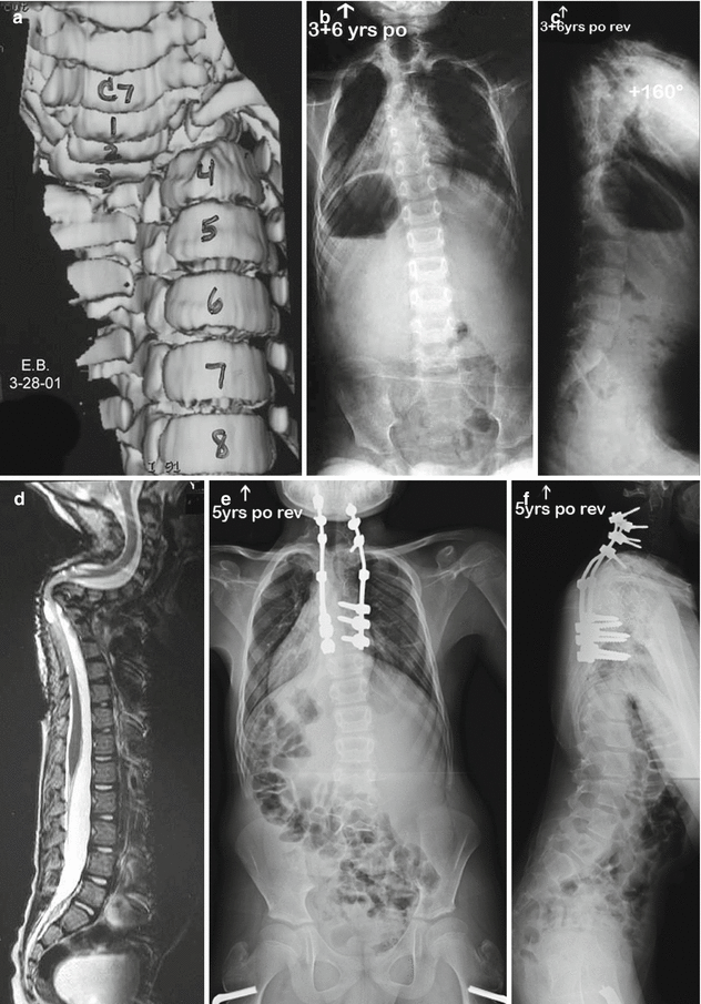
Fig. 36.2
Patient is a 2-year-old boy who presented with a congenital dislocation of T3/T4 as seen on (a) his preoperative coronal 3D CT scan. At three years of age, he underwent in situ fusion with subsequent deformity progression and myelopathy. (b, c) At the age of 5 (3 + 6-years postoperative), his coronal and sagittal x-rays somewhat hid his severe deformity. (d) However, on his sagittal MRI scan, the angular deformity of the cervicothoracic region with tenting of the spinal cord is obvious causing myelopathy. (e, f) He underwent a posterior revision reconstruction and a VCR of T3 and T4 for spinal cord decompression with ultimate midcervical to midthoracic instrumentation and fusion for stabilization of his deformity. His myelopathy completely resolved following decompression
36.2.3 Genetic Syndromes
Unfortunately, many genetic syndromes have musculoskeletal conditions involving progressive spinal deformity. Some of the more common are those involving the connective tissues, such as Marfan disease and Ehlers-Danlos syndrome. In a young patient, this becomes a detrimental combination involving a large deformity and very lax connective tissues. Growth modulation techniques should be utilized in the highly immature patient, but again, once patients are nearer to age 10, an apical if not complete spinal fusion should be considered. If left untreated, these patients may ultimately develop severe, life-threatening deformities because of their poor connective tissue; hence, surgical intervention can produce a secure and stable spinal column.
36.2.4 Neuromuscular Disorders
There is a spectrum of diagnoses involved in neuromuscular spinal deformity. Unfortunately, most patients do not benefit from a prophylactic orthosis, especially when their deformity reaches severe Cobb measurements of over 90–100°. In addition, many of them are not very healthy and thus may not be able to tolerate repeated surgical intervention necessary with treatments such as growing rods. Thus, entertaining a spinal fusion, especially during the EOS years, may be appropriate. One diagnostic category where this is certainly advantageous is with spinal muscular atrophy. These patients may develop a very severe deformity at a very young age and often are not very healthy from a pulmonary standpoint. Thus, it is doubly troubling as not only are they not healthy enough to tolerate an anterior procedure, they are also young enough to have a high risk of crankshaft phenomenon occurring if they undergo a posterior-only arthrodesis. However, this has been our approach, and a minimum 5-year postoperative review of these patients has shown reasonable radiographic and clinical results following early intervention for progressive, severe spinal muscular atrophy scoliosis [6].
Another category where spinal fusion has been performed at a very young age is congenital kyphotic deformities of the lumbar spine and those patients with a high-level myelomeningocele. Once a lumbar congenital kyphosis reaches over 100° or so, nothing advantageous is going to happen to that region of the spine; hence, reconstruction via a kyphectomy and spinal fusion is indicated. Most surgeons would attempt to “grow” the middle and upper thoracic spine with implants such as a Luque trolley or other growth-guided techniques such as Shilla, if at all possible, to allow further thoracic growth despite the thoracolumbar and lumbar fusion. It is particularly advantageous to avoid the mid and lower thoracic lordotic deformity that often becomes fixed in response to a progressive lumbar kyphosis below.
Most patients with cerebral palsy with a scoliosis deformity can be observed and braced or undergo wheelchair modification during the EOS years, in order to avoid spinal fusion until adolescence. However, once again, in those with severe, relentlessly progressive deformities greater than 100–125°, continued observation seems inappropriate when so much growth remains. Obviously, if they are healthy enough to undergo multiple surgical procedures, some type of posterior growing rod construct may be advisable. However, many of these patients are frail and debilitated, and a better option might be a posterior spinal fusion utilizing intraoperative halo-femoral traction to maximize correction and sitting balance while minimizing complications [7]. It is essential to explain to caregivers the full ramifications of a spinal fusion at such a young age.
Related posts:
 Anesthesia and Postoperative Management of Spinal Deformity Surgery in Growing Children
Regulatory Policies Regarding Pediatric Spinal Devices
Imaging of the Growing Spine
Orthotic Management for Early Onset Scoliosis
Growth Modulation Techniques: Titanium Clip-Screw Implant System (HemiBridge)
Single and Dual Traditional Growing Rods
Anesthesia and Postoperative Management of Spinal Deformity Surgery in Growing Children
Regulatory Policies Regarding Pediatric Spinal Devices
Imaging of the Growing Spine
Orthotic Management for Early Onset Scoliosis
Growth Modulation Techniques: Titanium Clip-Screw Implant System (HemiBridge)
Single and Dual Traditional Growing Rods
Stay updated, free articles. Join our Telegram channel

Full access? Get Clinical Tree


