Fig. 11.1
The anterior formation defect (a) is accompanied with fused lamina on the posterior spine (b)
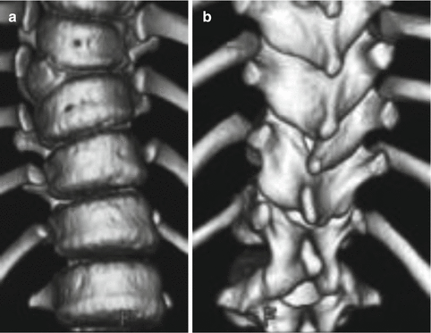
Fig. 11.2
The level of the anterior (a) and the posterior (b) vertebral malformation is discordant
Kawakami et al. [35] indicated that failure of formation in Winter’s classification includes those with segmentation failure such as nonsegmented hemivertebra, although those can be classified into failure of segmentation or mixed type. The report classified congenital spinal deformity into the solitary simple, multiple simple, multiple complex, and pure type of segmentation defect based on these three-dimensional morphological findings. This new classification is based on the concept that vertebral anomalies in failure of segmentation do not have any type of abnormal vertebrae with the characteristics of formation failure (Fig. 11.3). Although this classification is slightly more complicated, it demonstrates necessity of a preoperative three-dimensional morphological analysis for determining the optimal surgical strategy for treating congenital vertebral anomalies.
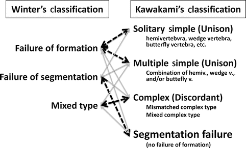
Fig. 11.3
The new classification of congenital spinal malformation by Kawakami et al. and Winter et al.
Winter et al. Classification [60]
I.
Unclassifiable: There is a collection of many types of segmentation defects. There is no dominating type.
II.
Fusion of ribs.
III.
Unilateral failure of formation of a vertebra, partial: this produces a wedge or trapezoid-shaped vertebra. A vestigial pedicle may be present.
IV.
Unilateral failure of formation of a vertebra, complete: this produces a hemivertebra.
V.
Bilateral failure of segmentation: this refers to the condition in which there is absence of the disc space between adjacent vertebral bodies.
VI.
Unilateral failure of segmentation: this produces an unsegmented bar and may involve two or more vertebrae and only the bodies or only the posterior elements.
Kawakami et al. Classification [35]
Type 1. Solitary simple type: there is only one abnormal vertebra in the entire curve.
Type 2. Multiple simple type: there are multiple abnormal vertebrae with a consistent anterior and posterior structure.
Type 3. Multiple complex type: there are multiple abnormal vertebrae with a combination of formation and segmentation defects with or without discordancy.
Type 4. Segmentation failure type: there are multiple abnormal vertebrae without any type of formation failure.
11.4 Natural History
Longitudinal spine growth comes from superior and inferior end plates. Curve progression is caused by unbalanced growth of one side of the spine. Well-formed and normal appearing discs suggest healthy growth plates and potential for asymmetric growth. On the other hand, the presence of bar or fused ribs is a sign of restricted growth on this side and may cause progressive deformities [29]. Therefore, the progression of CS depends on the type of the anomaly. The location of vertebral malformation and the growth potential of the patient are the other two most important factors in predicting the deterioration potential of the curve.
The two most important natural history studies were published by Winter et al. [60] in 1968 and McMaster and Ohtsuka [41] in 1982. Winter et al. [60] followed 234 patients with CS and found that thoracic and thoracolumbar curves progressed more than cervico-thoracic and lumbar curves did. A mild cervico-thoracic curve might cause serious cosmetic deformity because of head tilt, prominence of the neck, and dropping of one shoulder. It was also found that the rate of progression was not related to the severity of the curve, since some of the mild curves progressed more rapidly than the severe ones. Progression was most likely to occur when there were multiple unilateral anomalies in the thoracic spine. It was also reported that most severe deterioration of the curve was seen during preadolescence and infancy period. McMaster and Ohtsuka [41] followed 216 patients for a mean of 5.1 years and reported that the rate of curve deterioration was found to depend on both the level and the type of malformation. For each type of deformity, the deterioration of the curve was less severe in upper thoracic regions, more severe in mid-thoracic region, and worst in thoracolumbar region. Block vertebra and bilateral failure of segmentation are the most benign forms of anomaly, and the progression rate is less than 2° per year. Wedge vertebra, hemivertebra, and unilateral bar cause more severe deformities, respectively. A unilateral bar and contralateral hemivertebra were the most severe anomalies and have a progression rate of 5–10° per year. On the other hand, Winter et al. [62] later reported spontaneous improvement of scoliosis in seven patients with a hemivertebra in a review of 1250 with congenital spinal deformity. Therefore, predicting curve progression is still difficult. This may be due to the variety of not only vertebral body morphology but also posterior structure. Further analysis of the three-dimensional vertebral morphology using computed tomography (CT) and magnetic resonance imaging (MRI) images may reveal more precise determination of the natural history of each type of vertebral anomaly.
11.5 Patient Evaluation
The evaluation of a patient with CS focuses on physical examination including detailed spine and neurological examination, radiographic evaluation, and investigation of associated anomalies.
11.5.1 Physical Examination
Since the spinal growth is a major concern in CS, physical examination should start with recording the sitting and standing height and weight. The growth of the child should be monitored, as there is a close relationship between growth and curve progression, as discussed in natural history part.
CS may cause spinal imbalance; therefore, sagittal and coronal spinal imbalance should be recorded. Spinal balance in sagittal and coronal plane, and pelvic balance, head tilt, and shoulder balance must be keenly recorded. The rigidity of the curve is assessed.
Rib cage deformities can be seen with vertebral malformation; therefore, any anomaly of rib cage should be recorded. Inspiratory and expiratory capacity of lungs should be evaluated by pulmonary function tests to detect any restrictive lung disease.
A detailed neurological examination including muscle forces, sensation of the skin, abdominal and deep tendon reflexes should be recorded to rule out any spinal dysraphism. Patient’s back should be examined carefully for any hair patches, lipomata, dimples, and abnormal pigmentation, which can be the first signs for intraspinal pathology. Physical findings such as asymmetrical calves, cavus feet, clubfeet, and vertical talus can also be the manifestations of spinal dysraphism, so a detailed lower extremity examination is mandatory.
11.5.2 Imaging
Appropriate imaging techniques should be used during patient evaluation in order to define the pathologic anatomy, classify the malformation, and make a logical surgery plan.
Routine radiographs are essential to evaluate the deformity. Radiographs in infants can be taken supine. As the child becomes older and can stand independently, standing posterior–anterior (PA) and lateral views should be obtained [29]. Measurement of Cobb angle is often more difficult in patients with CS, because of the distorted end plates and the malformed pedicles. However, with high-quality radiographs, it is possible to determine the type of the malformation, the magnitude of the curve, and the growth potential of the vertebral anomaly. It is also a reliable method for follow-up of curve progression [18].
In a study investigating intra- and interobserver variability in measurement of the Cobb angle in CS, Loder et al. [37] found a variability of ±9.6° and ±11.8°, respectively. According to the authors, to ensure with 95 % confidence that the increase in the curve is not due to error of measurement, at least 23° of change is necessary. Facanha-Filho et al. [18] stated that the variability in Loder et al. study was very high and performed another study to assess variability in measurement of the Cobb angle. They found a mean intraobserver variance with an average of 2.8°, and the interobserver variance was 3.35°.
It is possible to estimate the growth potential of malformed vertebrae from disc spaces and their relative sizes on direct radiographs. If they are narrow and poorly defined, they do not have much growth potential. On the other hand, visible, wide, normal appearing discs associate a high potential for growth and curve progression. Even though the conventional method of evaluating CS is direct roentgenograms, these images can be difficult to interpret in patients with small size, in overlying structures obscuring the deformity, and in complex deformities. In patients who are candidates for surgery, more detailed imaging techniques are necessary.
New improvements in computed tomography (CT) and magnetic resonance imaging (MRI) technology made both of these modalities indispensable, especially in patients undergoing spinal stabilization and with complex deformities.
Three-dimensional computed tomography (3D-CT) is the best modality for defining the osseous anomalies and their relationships [46]. It is mostly recommended for complex deformities but not for routine observation or serial follow-up. Hedequist et al. [28] compared the findings in direct radiogram and 3D-CT of patients with CS with the findings in surgery. In all patients, anterior and posterior anatomy correlated with the CT findings. It is clear that a three-dimensional analysis of abnormal vertebrae can demonstrate the relationship between the anterior and posterior structures. These findings are absolutely necessary to evaluate even a single hemivertebra, whether it is a discordant type or not, to ensure that the patient undergoes the appropriate level of surgery [35] (Fig. 11.3). On the other hand, it should be kept in mind that serial X-ray and frequent CT evaluations loads significant amount of radiation to those patients with CS and every effort should be spent to decrease the unnecessary radiographic evaluations.
EOS (EOS Imaging, Paris, France) is a novel low-dose biplanar digital radiographic imaging system that can scan the patient in a standing position in two orthogonal planes. Although it has been shown that the EOS system can be reliably used in adolescent idiopathic scoliosis and limb length measurements, its reliability has been questioned in CS where each individual patient shows different curve and malformation patterns [17, 30]. The main advantage of EOS is that it decreases radiation exposure by up to 85 %.
MRI is the standard diagnostic tool for the assessment of intraspinal pathology [11]. Specific indications for MRI include the presence of neurological signs such as weakness, sensory loss, bowel or bladder dysfunction, a skin abnormality over the spine like dimple, hairy patch or nevus, leg or back pain, lumbosacral kyphosis, interpedicular widening. MRI is also very important in any patient undergoing spinal correction and stabilization. In addition, MRI may become a key modality for the assessment of natural history of congenital vertebral anomalies, particularly hemivertebrae because it also demonstrates various types of congenital disc and tissue anomalies [44] (Fig. 11.4).
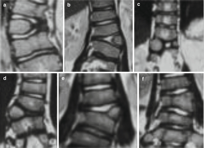
Fig. 11.4
(a–f) Magnetic resonance imaging (MRI) of different types of hemivertebras and the discs around malformed segments
Since the genitourinary system abnormalities are found in 18–40 % of patients with CS, screening renal ultrasonography is recommended to all patients [15, 39]. The incidence of congenital heart disease in patients with CVM was found to be 26 % in a recent study of Basu et al. [8] revealing the high importance of detailed cardiac examination and an echocardiography for a thorough examination.
11.5.3 Associated Anomalies
The development of vertebral column is closely associated with the development of the spinal cord; therefore, the neural and vertebral malformations often coexist. These malformations may cause neurological findings. However, absence of any neurological finding does not rule out intraspinal pathology [29]. Mesoderm, which is responsible for the formation of vertebra, is also responsible for the formation of urogenital, pulmonary, and cardiac systems. Malformation of these systems can also accompany congenital vertebral malformations [33]. Therefore, systemic evaluation of the patients with proper imaging techniques is mandatory.
In a study using direct radiographs and myelograms, McMaster et al. [42] found intraspinal pathology in 18.3 % of 251 CS patients. When MRI was used as diagnostic tool for evaluation of the CS, neural axis abnormalities increased to 30–38 % [11, 48]. Bollini et al. studied the relationship of the level of the hemivertebra (HV) and the incidence of the intraspinal pathology. They showed that the incidence of intraspinal pathology is higher in patients with HVs located at lumbosacral region than any other part of the spine (33 % for lumbosacral, 13 % for lumbar, and 10 % for thoracic HV). They were unable to show any relationship between the intraspinal or other visceral pathologies and the type (segmented-semisegmented), number (single-multiple), side (right-left) of the HV, or gender of the patient [10].
The most common malformation of spinal cord is diastematomyelia (split cord), which is defined as partial or complete split of spinal cord or cauda equina with a bony or fibrous spur [42]. Diastematomyelia is found in approximately 20 % of patients with congenital scoliosis [27]. In patients with diastematomyelia, the normal movement of spinal cord is restricted. The spinal cord stretches while the spinal column grows longitudinally. Any corrective manipulation of spine may cause more stretching of the cord, and this may result in neurological deterioration. Therefore, it is very important to evaluate the whole spine before any corrective surgery. Other congenital intraspinal anomalies associated with CVM are epidermoid cysts, dermoid cysts, neuroenteric cysts, tethered spinal cord, lipomas, and teratomas [42] (see Chap. 15).
Evidence from a number of retrospective studies shows diminished pulmonary function in patients with CS. Because of the complex interconnections between spine, sternum, and ribs, the displacement and rotation of the vertebrae in scoliosis have profound effects on the shape of the thorax. In a retrospective study, 192 (50.3 %) of 382 patients were reported to have rib anomalies. Missing rib was the most common anomaly among others. The anomalies were located more frequently on the concave side and mostly associated with thoracic and thoracolumbar CVMs [63].
Individuals with CS and chest wall deformities are believed to have a thoracic deformity that limits lung growth and rib deformities leading to thoracic instability and alteration in respiratory mechanics. The expansion of the thoracic cavity is limited as the movement of the ribs is impeded, which in turn decreases chest wall compliance and makes breathing significantly harder despite the absence of any lung disease. Altered development and morphology in patients with scoliosis can lead to measurable changes in lung function most consistent with restrictive lung defect.
Renal system abnormalities may be found in 18–40 % of patients with CVM. Anomalies may affect the kidneys, ureters, bladder, and urethra. Unilateral renal agenesis, duplicated kidneys, and ureteral obstruction are the most common renal abnormalities associated with congenital scoliosis [15, 39].
Congenital heart disease is present in 10–26 % of patients. Atrial and ventricular septal defects are the most common cardiac abnormalities. More complex cardiac malformations like tetralogy of Fallot and transposition of great vessels can also be seen in CS patients [8, 49].
Musculoskeletal anomalies like clubfeet, Sprengel’s deformity, Klippel-Feil syndrome, developmental dysplasia of the hip may all be seen in these patients [29].
11.6 Treatment Alternatives
Once the CS is diagnosed, it is very important to note the patient’s age and spinal balance and to classify the anomaly. Treatment should be started regardless of age, if the patient has CVM with high potential to deteriorate such as unilateral hemivertebra and contralateral unsegmented bar. Patients with anomalies less inclined to deteriorate should be carefully followed with serial radiographs, and Cobb angle measurement of their curves should be obtained at each visit to detect any progression.
There are many treatment alternatives for CS. The age of the patient, type of anomaly, limitations of the surgeon, and surgery room should be considered to choose the most appropriate way of treatment.
11.6.1 Observation
Patients with balanced spine and vertebral malformations, less prone to deteriorate like hemimetameric shift or block vertebra, can be followed with serial plain radiographs at 4- to 6-month intervals. It is important to assess the spinal balance of the patient and the Cobb angle measurements of the curves. Most recent radiographs should be compared with the earliest radiographs of the patient in order to detect any progression.
11.6.2 Bracing and Casting
Short and rigid curves rarely respond to brace treatment. Bracing can be considered for long, flexible curves and for compensatory curves, which are located proximal or distal to anomalous segment. Serial casting, which is an effective treatment modality in early-onset idiopathic scoliosis, can also be used in very young patients with CS until surgical procedures could be applied. Demirkiran et al. recently showed that the serial casting can effectively control both the main and the compensatory curves while providing longitudinal spine growth in 11 patients with CS [14].
11.6.3 Surgery
Surgical techniques applied in CS will be discussed in detail in consecutive chapters, so only brief summaries regarding the surgical alternatives will be mentioned here. The reader is advised to refer to specific chapters for technical details (Chaps. 30, 31, 32, 33, 39, and 40).
Related posts:
 Anesthesia and Postoperative Management of Spinal Deformity Surgery in Growing Children
Regulatory Policies Regarding Pediatric Spinal Devices
Imaging of the Growing Spine
Orthotic Management for Early Onset Scoliosis
What Can We Learn About Ribs and Vertebra Growth from an Osteological Collection?
Single and Dual Traditional Growing Rods
Anesthesia and Postoperative Management of Spinal Deformity Surgery in Growing Children
Regulatory Policies Regarding Pediatric Spinal Devices
Imaging of the Growing Spine
Orthotic Management for Early Onset Scoliosis
What Can We Learn About Ribs and Vertebra Growth from an Osteological Collection?
Single and Dual Traditional Growing Rods
Stay updated, free articles. Join our Telegram channel

Full access? Get Clinical Tree


