Fig. 46.1
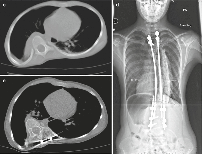
(a, b) Preoperative radiographs of a 5-year-old female with neglected 75° lordoscoliosis and Marfan syndrome. T1–12 length = 16.5 cm. (c) Apical CT of thorax. Significant spine penetration into the convex hemithorax, as well as narrowing of the ventral-dorsal lung space due to lordosis, is seen. (d) AP radiograph after 3 years of distraction-based growing rod instrumentation. T1–12 length = 23.6 cm, scoliosis reduced to 46°. (e) Apical CT after 3 years of distraction treatment. In spite of significant length gain, there is little change in the spinal penetration, apical rotation, or ventral-dorsal lung space
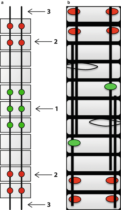
Techniques to control apical deformity, and at the same time avoid the serial lengthening procedures required by “traditional” growing rod instrumentation, have been added to the EOS treatment armamentarium. Growth-guidance procedures [12, 14], described in detail elsewhere in this textbook, provide direct control of the apical deformity by vigorous correction of apical segments, and then by extending instrumentation to the end vertebrae with non-constrained, “sliding” anchors as part of the construct, permit continued spinal growth along a path dictated by the non-constrained longitudinal members of the construct . In the “Shilla” technique, growth occurs outside the fused apical segments, elongating as the spine grows away from the apex (Fig. 46.2a). In the modern “trolley” technique, growth occurs in the non-fused but “guided” apical segments between the end-vertebral anchors which are fused in place (see Fig. 46.2b). Both methods enjoy the theoretical advantage over conventional growing rod instrumentation (GRI) of not requiring serial surgical lengthening procedures in order to accomplish deformity control while permitting continued spine growth.


Fig. 46.2
(a) Shilla construct. Apical anchors (1) are formally locked to the rods after vigorous short-segment deformity correction. End-vertebral anchors (2) are “sliding” anchors, captured by the set screws but not locked to the rods, permitting “guided” growth of the non-fused segments within the construct. Extra length of rods (3) outside the end vertebrae must be available in order for guided growth to occur (b) modern “trolley” construct. End-vertebral anchors (red) are fused into place to provide stable anchoring points for the rods. Sliding anchors (green) and sublaminar wires which capture the overlapping rods as well as the spine segment control the apical deformity by cantilever correction as well as permit longitudinal growth with the rods sliding apart (a: Courtesy R.E. McCarthy, MD; b: Courtesy J.A. Ouellet, MD)
Recent reports [2, 12, 13] have elucidated the limitations of the Shilla method, namely, technical complications requiring frequent unplanned revisions while achieving less length gain and deformity reduction than matched cases treated by conventional GRI procedures. Results of the modern trolley are too limited to make outcome comparisons with conventional methods valid. However, the importance of apical control of EOS deformity has been reaffirmed in a series of cases described in this report, combining the use of apical segment anchors with conventional GRI technique to achieve axial plane correction progressively with elongation of the spine.
46.2 Rationale
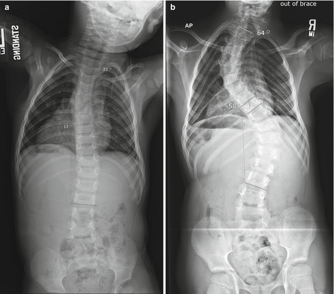
The concept of the windswept thorax was originally introduced by Dubousset referring to the penetration of the lordoscoliotic apex into the convex hemithorax [8], narrowing the space for lung volume – already narrowed by the rotatory rib deformity – to the most peripheral area between the spine and the chest wall (Fig. 46.1c). In severe cases, the space remaining for convex lung volume is reduced to a narrow sliver as the spine lateral translation into the convex hemithorax squeezes the lung against the chest wall. The term “collapsing parasol” has been used to describe the relative vertical position, or “drooping,” of ribs in primarily hypotonic neuromuscular patients [7], but this description can certainly be applied to the convex rib deformity in any form of EOS (Fig. 46.3a, b). Because the anatomic volume of the hemithorax is so diminished by the combined apical rotation and translation, direct control of this portion of the spine is mandatory if more effective corrective measures to reverse the windswept deformity are to be undertaken. The use of apical pedicle screw anchors, for example, placed only on the convexity to allow further concave growth, permits direct derotation and translation of the apex toward the concave hemithorax (Fig. 46.4a, b). By fusing only the apical segment to stabilize the convex anchors, and then progressively recorrecting the lateral translation toward the concavity by in situ rod contouring at each subsequent scheduled lengthening, the goal of controlling – indeed improving – deformity while permitting or driving growth, fundamental to the management of EOS, can be achieved [10]. The procedure described here combines progressive apical correction with serial lengthening – the best effects of traditional GRI and the growth-guidance concepts.


Fig. 46.3


(a) A 20-month-old male with syndromic, hypotonic scoliosis. First standing radiograph. Rib morphology is unremarkable. (b) Age 7, spine deformity has progressed along with apparent chest wall narrowing and “drooping” of the ribs (“closed parasol”), possibly due to casting and brace treatment
Stay updated, free articles. Join our Telegram channel

Full access? Get Clinical Tree


