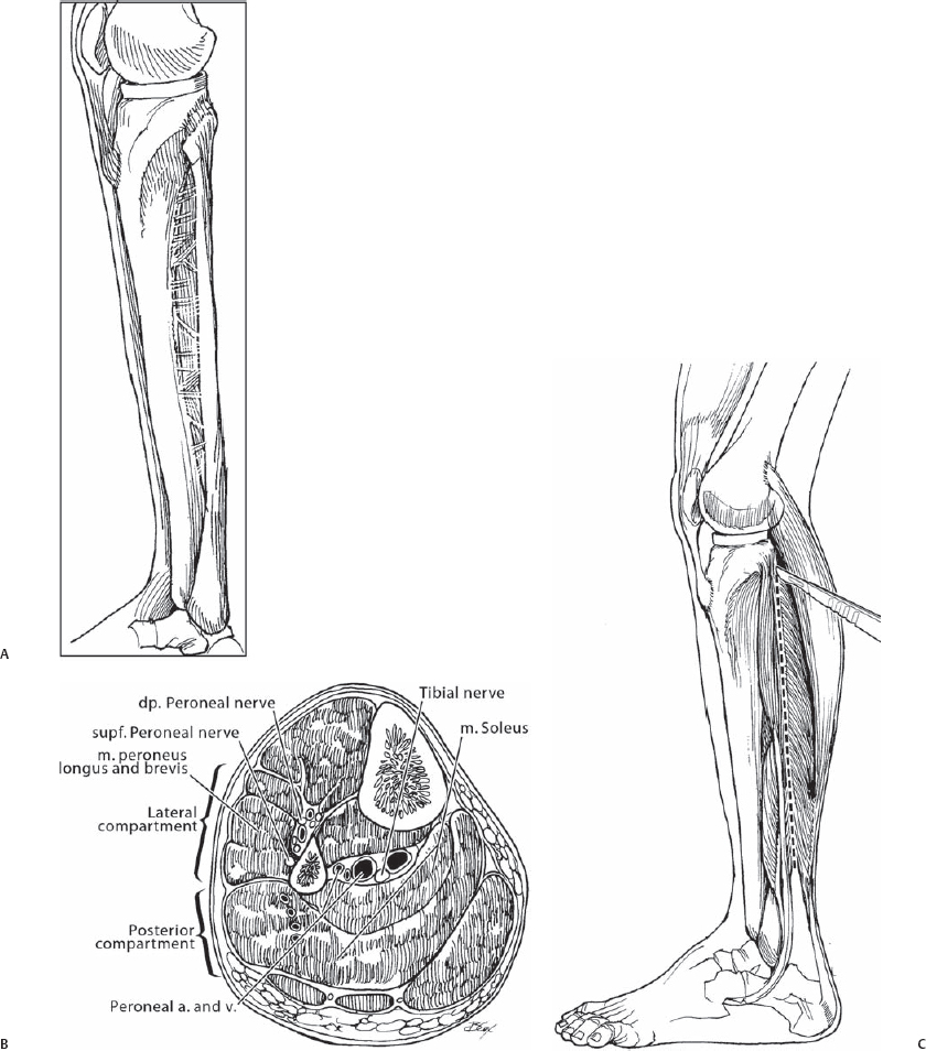
71 Joshua D. Auerbach and Kingsley R. Chin To harvest autologous fibula or rib bone graft for spinal reconstructive procedures while minimizing donor-site morbidity. Although autologous fibula and rib strut grafts are not routinely used for spinal reconstructive surgeries, they remain excellent options for those procedures that require long strut grafts. Autologous graft harvesting from the fibula or rib carries minimal added infection risk, achieves favorable rates of fusion, provides a source of vascularized cortical bone, and can be safely performed with minimal donor-site morbidity. Autologous fibular strut grafts have superior compressive strength compared with iliac crest and rib grafts. Safe harvest of autologous fibula or rib grafting for use in spinal reconstructive procedures. Multilevel cervical corpectomies, posterior cervical reconstruction, posterior occipitocervical fusion, anterior thoracic spinal fusion and reconstruction for tuberculosis spondylitis, infection, neoplasm, and progressive kyphosis. In situations at high risk for delayed union or nonunion, such as multilevel fusions, thoracic/thoracolumbar kyphosis, or in tumor patients receiving perioperative radiation, vascularized fibular or rib grafts may be indicated to provide a solid initial biomechanical construct and facilitate a quicker time to fusion. If a vascularized fibular graft is considered, it is recommended to obtain preoperative arteriography to evaluate for anomalous vascularity or previous vascular injury of the fibular pedicle graft. The fibula is a straight cortical bone that can provide a graft up to 26 cm in length. The vascular pedicle, which may be from 1 to 5 cm in length, is composed of the peroneal artery (1.5 to 2.5 mm in diameter) and its accompanying two vena comitantes (2 to 3 mm in diameter). The rib is a curved, flexible bone that can provide a graft up to 30 cm in length. The shape of the graft can be modified by the choice of the rib and section of rib excised (from the flatter posterior portion to the curved anterior portion) to optimize contact between the graft and recipient bed. Its vascular pedicle, which may be from 3 to 5 cm in length, consists of the posterior intercostal artery (1.5 to 2 mm in diameter) and single intercostal vein (1.2 to 2.5 mm in diameter). The fibular graft site can be approached using either a posterior or lateral approach. The lateral approach is preferred, as it is simpler and quicker and can be done simultaneously with the majority of the indicated primary procedures. With the patient supine, a sandbag or roll is placed underneath the ipsilateral buttock for optimal visualization of the lateral leg. A tourniquet is applied to the thigh to maintain a bloodless field. General anesthetic is typically used, with or without supplementation with local long-acting anesthetic. The patient can be placed in either the prone position if a posterior spinal procedure is anticipated, or in the lateral decubitus position with the graft side up, which allows for visualization of the rib along its entire course. In the lateral decubitus position, the ipsilateral arm is elevated anteriorly and cephalad away from the ribs and the table flexed at the midthoracic region to increase the intercostal space. The table is tilted anteriorly to allow the ipsilateral lung to fall anteriorly. A double-lumen tube is used to allow single lung ventilation. General anesthesia is typically used, with or without supplementation with local long-acting anesthetic. Obese patients may increase the difficulty of localizing the anatomy, especially the middle ribs. Have a low threshold to use fluoroscopy or plain radiographs to identify the correct rib. Excessive bleeding may occur after releasing the tourniquet, requiring reinflation and a vascular consult. To avoid damage to the peroneal nerve, proximal fibular osteotomy should be performed at the junction of the middle and distal thirds of the fibula, whereas ankle instability can be safely avoided by staying 10 cm above the ankle joint for the distal fibular osteotomy. A straight lateral approach to the midportion of the fibula is used. Dissection is initially performed between the posterior and lateral compartments of the leg. Incise the superficial fascia overlying the peroneus longus and soleus muscles (Fig. 71.1), and continue to dissect in a plane posterior to the peroneus longus and anterior to the soleus muscle. The peroneal vessels lie just deep to the soleus and are almost in contact with the fibula. After identification and protection of the vessels, incise the fibular origin of the soleus muscle and retract posteriorly (Fig. 71.2). Identify and protect the superficial peroneal nerve in the proximal aspect of the incision. Attention is then turned to the distal aspect of the fibula, where the interval between the peroneal muscles and the more posteriorly located flexor hallucis longus is identified. The peroneal vessels travel within the substance of the flexor hal-lucis longus muscle and are therefore protected with careful retraction. For anterior exposure, retract the peroneal muscles anteriorly and dissect them off the fibula in an extraperiosteal plane. Continue extraperi-osteal dissection distally, being sure to protect the anterior tibial artery and deep peroneal nerve anteriorly (Fig. 71.3). If no vascularized graft is warranted, however, dissection can be performed in a subperiosteal plane to maximize protection of the neurovascular structures. A Gigli saw is used to osteotomize the fibula with careful retraction of the peroneal vessels posteriorly, and the superficial and deep peroneal nerves and anterior tibial artery anteriorly.
Autologous Fibula and Rib Harvesting
Description
Key Principles
Expectations
Indications
Contraindications
Special Considerations
Special Instructions, Position, and Anesthesia
Fibula
Rib
Tips, Pearls, and Lessons Learned
Difficulties Encountered
Key Procedural Steps
Fibular Harvest

Stay updated, free articles. Join our Telegram channel

Full access? Get Clinical Tree


