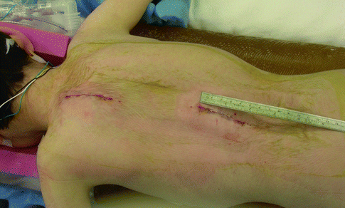
Fig. 48.1
Dr. Robert Campbell holding Clay Skaggs, 2001, Los Angeles (Reproduced with permission of Children’s Orthopaedic Center, Los Angeles)
There are many potential benefits for using ribs as anchors (Table 48.1), the first of which is that the rib attachments may allow for motion preservation. The ribs are attached to the spine via the costotransverse joints (neck of rib to anterior portion of transverse process) and the costovertebral joints (head of rib to vertebral body). The costovertebral joints constitute a series of gliding or arthrodial joints formed by the articulation of the rib head with the facet on the contiguous vertebrae. Ribs 1, 10, 11, and 12 articulate with single vertebral bodies; the remaining ribs attach to two vertebrae [4] (Fig. 48.2a, b). These joints permit a gliding motion. During normal respiration, there is just over 10° of bucket-handle motion of the ribs relative to the spine [5]. In addition, the interface of a hook on the rib is not rigid and allows for some “slop.” Yamaguchi et al. reviewed a series of 176 patients treated with growing rods. In this series, proximal rib anchors were protective against rod breakage with a 77 % decreased incidence compared to proximal spine anchors [6]. Additionally, rib hooks may have an advantage in preventing anchor failure. Akbarnia et al. performed the first study to evaluate the properties of rib hooks used as an upper growing rod foundation in a porcine cadaver model [7]. They showed that rib hooks had a significantly higher load to failure than transverse process–lamina hook pairs and lamina hook–lamina hook pairs. Rib hooks had a higher load to failure than pedicle screws but this was not significant.
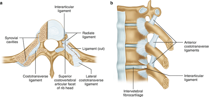
Table 48.1
Possible benefits of spine implants as rib anchors
Motion preservation |
No dissection of spine |
Good soft tissue coverage |
No special equipment, training, or institutional approval needed |
Load sharing over multiple ribs |
No fusion of the upper thoracic spine, which is important for pulmonary development |

Fig. 48.2
(a, b) Demonstration of the costotransverse and costovertebral joints
In contrast, traditional growing rods attached to the spine permit little motion of the vertebrae within the construct. As with any diarthrodial joint, prolonged immobilization decreases motion and can lead to spontaneous fusion. It is a common experience upon converting growing rods to a final fusion construct to discover spontaneous fusion of most, if not all, of the vertebrae within the construct. Sankar et al. substantiated this theory demonstrating in patients treated with dual growing rods that over time there was less increase in T1 to S1 distance with each subsequent growing rod lengthening [8]. This “law of diminishing returns” is a particular concern when growing rods are placed in young children, i.e., if dual rods are placed in a 2-year-old, the spine may be fused by age 7. In theory, the use of the ribs for anchor points will permit motion between vertebrae and prevent or delay spontaneous fusion. This technique is too new to have adequately tested this theory.
Another advantage of using ribs as anchors is to stay out of the spine and preserve a virgin spine for future surgeries. While traditional growing rods aim for a fusion at the top of the construct, methods that attempt nonfusion, such as Luque-Trolley (sublaminar wires and rods without fusion), have been shown to cause fusion in 100 % of patients in one series [9]. Fusion in the upper thoracic spine (T1–T3) in young children is particularly harmful to long-term pulmonary function, so avoiding fusion in this region may be an important benefit of this technique [10].
It has been shown that children with thoracic insufficiency are nutritionally depleted, with 79 % being below the fifth percentile for weight [11]. Soft tissue coverage of traditional spine implants can be challenging in this population. A further advantage of rib anchors is that they tend to have good soft tissue coverage, as they are located deep to the rhomboids and trapezius, in a valley between the more prominent spine and scapula (Fig. 48.3).
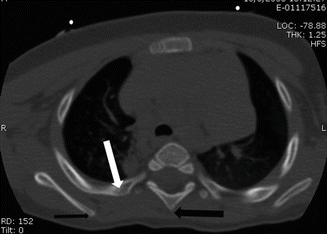

Fig. 48.3
CT of the chest. Note that the attachment point of the rib anchor (white arrow) is in a trough with good soft tissue coverage between the spinous process (thick black arrow) and scapula (thin black arrow) (Reproduced with permission of Children’s Orthopaedic Center, Los Angeles)
A significant practical advantage of using traditional spine implants on ribs rather than VEPTR is that no special equipment is needed. Hooks that fit the ribs are readily available on all spine implant systems. There are, thus, no special equipment needs and no special training needed for the surgeon or operating room staff. In addition, there is no need for institutional or research approval as traditional spine implant hooks are FDA approved. A particular advantage over the original VEPTR design is that load sharing over multiple ribs is quite simple (Fig. 48.4).
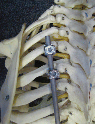

Fig. 48.4
With hooks of standard spine implants, multiple ribs may be engaged to share the load over multiple ribs. Note the hooks are immediately adjacent to the transverse process (Reproduced with permission of Children’s Orthopaedic Center, Los Angeles)
48.2 Indications
The indications for use of ribs as anchors in growing systems continue to evolve. This remains an area where further investigation is needed and a particular area of uncertainty even among experienced early onset scoliosis surgeons [3]. The argument may be made to use rib attachments in children under the age of 5 years, as they would be expected to have growing implants for at least 5 years, and rib attachments may decrease the risk of spontaneous fusion as discussed earlier. Another indication is when there is already a substantial fusion of the mid thoracic spine present (whether from previous surgery or congenital), and one wants to minimize further fusion in the upper thoracic spine. In cases of previous infection of growing implants, the ribs provide an area of new, uninfected tissue as salvage. Using rib anchors also allows one to avoid sites of previous surgery such as laminectomies.
Another indication for using ribs as anchors is in cases of significant cervicothoracic scoliosis and head tilt, in which the upper ribs are fused. This is discussed in detail near the end of the chapter.
48.3 Contraindications
Rib attachments tend to function poorly in cases of kyphosis as the ribs tend to pull backwards over time as the spine falls forward. In particular, upper thoracic kyphosis is poorly controlled with rib attachments, and this may be a time where traditional growing rods bent into kyphosis and attached cephalad to the region of kyphosis are more appropriate (Fig. 48.5a, b).
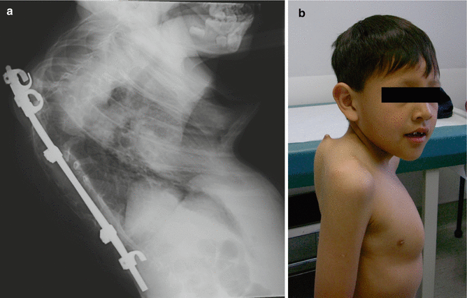

Fig. 48.5
(a) Lateral radiograph demonstrating progressive kyphosis despite rib implants. (b) Clinical photograph demonstrating rod coming through the patient’s skin (Reproduced with permission of Children’s Orthopaedic Center, Los Angeles)
48.4 Thoracotomy Generally Unnecessary
It is very uncommon that a formal thoracotomy is used with this technique. It has been shown that a thoracotomy in the treatment of scoliosis leads to a disruption of pulmonary function and is simply not needed to improve spinal and thoracic deformity in the great majority of cases [12]. Soft tissue or osseous release between ribs is rarely needed, except in the truly rare case of multiple rib fusions limiting thoracic expansion. It is natural that ribs are closer together in the concavity of a curve than the convexity, and that is not an indication for tissue destruction between the ribs. When the scoliosis is improved by distraction implants, the spaces between the ribs open up in a harmonious fashion (Fig. 48.6a, b). In contrast, distraction across a formal thoracotomy distracts between the two ribs at the site of the thoracotomy (Fig. 48.7). Any tissue lysis between ribs cannot help but leave scar tissue, which is less mobile and functional than virgin intercostal muscle.
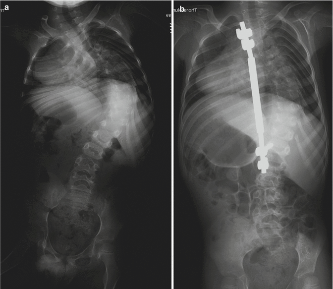
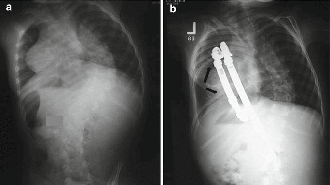

Fig. 48.6
(a) A preoperative AP radiograph of 91° scoliosis with ribs on the concave side appearing constricted. (b) Evidence of harmonious distraction of the ribs through distraction instrumentation without scapula elevation, thoracotomy, or any lysis of tissue between ribs (Reproduced with permission of Children’s Orthopaedic Center, Los Angeles)

Fig. 48.7
(a, b) Pre- and postoperative radiographs demonstrate large opening of two ribs at a thoracotomy site (between black arrows) and compression of rib spaces above the thoracotomy (Reproduced with permission of Children’s Orthopaedic Center, Los Angeles)
48.5 Surgical Technique
Neurologic monitoring should include both the upper and lower extremities. Joiner et al. have described the brachial plexus injuries that may occur in this setting [13]. Noted in this study were two cases in which the neurologic symptoms were present when adducted but resolved with the arms abducted in the typical positioning for a prone patient undergoing spine surgery. Consequently, positioning of the child with arms adducted is recommended to allow for accurate intraoperative monitoring (Fig. 48.8).
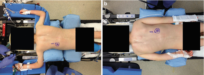

Fig. 48.8
Clinical photo showing typical prone position (a) and recommended adducted position (b)
The spine is approached through a midline skin incision as it is likely that this incision will be used for the final fusion in the future. Depending on the specifics of the surgery, a long midline incision or separate incisions at the top and bottom of the construct may be made. As the figure demonstrates, the top incision for the rib attachments is 4 cm in length (Fig. 48.9). The skin is undermined laterally past the transverse processes. The transverse processes are generally palpable as a point of resistance through the muscles. If there is any question, fluoroscopic imaging over a needle placed into bone clarifies the location. A combination of muscle splitting and cautery in a vertical incision should bring one quickly to the ribs with minimal blood loss.
