EEG-Based Brain-Computer Interfaces
Gert Pfurtscheller
Christa Neuper
INTRODUCTION AND BASIC PRINCIPLES
A relatively recent development in applied neurophysiology is an approach called EEG-based brain-computer interface (BCI). A BCI translates specific features, automatically extracted from EEG signals, into signals able to operate computer-controlled devices in order to assist patients who have highly compromised motor functions, such as tetrapalegic patients. This novel approach became possible due to advances both in methods of EEG analysis and in information technology, along with a better understanding of the psychophysiological correlates of certain EEG features. Therefore, it is interesting to take notice of the emerging field of direct brain-computer communication.
A BCI provides the brain with a new nonmuscular communication channel that can be used to convey messages and commands directly from the brain to the external world without using any muscle activity (1). Here, we expand this definition to emphasize that any BCI must have the following four components:
Direct: The signals must be recorded directly from the brain. If a device records signals after they pass through peripheral nerves or muscles, it is not a BCI.
Intentional control: At least one directly recordable brain signal, which can be intentionally modulated, must provide input to the BCI (electrical potentials, magnetic fields, or hemodynamic changes).
Real-time processing: The signal processing must occur online and yield a communication or control signal.
Feedback: The user must obtain feedback about the success or failure of his/her efforts to communicate or control.
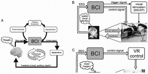
It follows from these definitions that each BCI is a closedloop system with two adaptive controllers: the user’s brain, which produces the signals and provides the input to the BCI; and the BCI itself, which analyses the brain signals and transforms them to a control signal as the BCI output (Fig. 57.1).
Any BCI contains components to extract features and classify (detect) EEG events. The goal of the feature extraction component is to find a suitable representation of the EEG signal that simplifies the subsequent classification or detection of specific patterns of electrical brain activity. That is, the signal features should encode the commands sent by the user but should not contain noise and other signal components that can impede the classification process. There is a variety of feature extraction methods used in current BCI systems. A nonexhaustive list of
these methods includes amplitude and band power measures, Hjorth parameters, autoregressive parameters, and wavelet coefficients (2, 3 and 4).
these methods includes amplitude and band power measures, Hjorth parameters, autoregressive parameters, and wavelet coefficients (2, 3 and 4).
The task of the classifier is to use the signal features provided by the feature extractor to assign the recorded samples of the signal to a given category of EEG patterns. In the simplest form, detection of an EEG pattern may be made, for instance, by means of a threshold method (5,6). More sophisticated classification algorithms of different EEG patterns depend on the use of linear or nonlinear classifiers (2,7,8).
The classifier output, which can be a simple on-off signal or a signal that encodes a number of different classes, is transformed into an appropriate signal that can then be used to control a variety of devices. For most current BCI systems, the output device is a computer screen and the desired output consists of the selection of certain targets. Advanced applications include controlling of spelling systems or other external apparatuses such as prosthetic devices and multimedia applications.
Feedback of performance is usually obtained by visualization of the classifier output on a computer screen or by presentation of an auditory, tactile, or visual feedback signal. Feedback is an integral part of the BCI system because the users observe, for example, selected letters or certain movements simultaneously with the brain responses they produce.
EEG PATTERNS USED AS INPUT FOR A BCI
The EEG is the most widely used brain signal in BCIs. Two types of changes can be extracted from the ongoing EEG signals:
Event-related potentials (ERPs) display time and phase-locked changes (evoked) to an externally or internally paced event. Evoked signals include slow cortical potential (SCP) changes, P300 components, and steady-state-evoked potentials (SSVEPs) (9).
Event-related changes in ongoing EEG activity in specific frequency bands. These changes are also time-locked but not phase-locked (induced). Event-related desynchronization (ERD) defines an amplitude (power) decrease of a rhythmic component, whereas event-related synchronization (ERS) characterizes an amplitude (power) increase (10).
Depending on the phenomena analyzed and classified, the following EEG-based BCI systems can be differentiated:
The SCP BCI: Beginning in 1979, Birbaumer and coworkers published a series of experiments demonstrating operant control of SCPs (see Ref. 11 for review). Operant conditioning is a learning process with the goal of the self-regulation of brain potentials (e.g., SCP shifts) or brain waves (e.g., sensorimotor rhythms) with the help of suitable feedback. This process does not require continuous feedback, but a reward for achieving the desired brain potential (wave) change is necessary. Operant conditioning was used in communication systems for completely paralyzed (locked-in) patients (12,13).
The P300 BCI: The P300 is the positive component of the evoked potential that may develop about 300 msec after an item is flashed. The user focuses on one flashing item while ignoring other stimuli. Whenever the target stimulus flashes, it yields a larger P300 than the other possible choices. P300 BCIs are typically used to spell (14, 15 and 16) but have been validated with other tasks such as control of a mobile robot (17) or a smart home (18).
The SSVEP BCI: Steady-state evoked potentials (SSEPs) occur when sensory stimuli are repetitively delivered rapidly enough that the relevant neuronal structures do not return to their resting states. In a BCI application, the user focuses on one of several stimuli, each of which flickers at a different rate and/or or phase. Gao et al. (19) described a BCI with 48 flickering lights and a high information transfer rate (ITR) of 68 bits/min. Like P300 BCIs, SSVEP BCIs require no training and can facilitate rapid communication (9,20,21). SSVEP BCIs have also recently expanded to tasks beyond spelling, such as controlling an avatar in a computer game (22, 23 and 24) or controlling an orthosis (25). Some BCI articles argued that the SSVEP can only be used for communication when users have some conscious control of eye muscles and is therefore not applicable for patients in the late stages of amyotrophic lateral sclerosis (ALS) (1,19). Later work showed that this assumption is incorrect; in some cases, SSVEP BCIs can function even when users do not shift gaze (9,26).
The ERD BCI: Brain rhythms can either display an event-related amplitude decrease or desynchronization or an event-related amplitude increase or synchronization (10). The term ERD BCI describes any BCI system that relies on the detection of amplitude changes in sensorimotor (mu and central beta rhythms) and/or other brain oscillations, also including short-lasting postimagery beta bursts (beta ERS, beta rebound) (8,27, 28 and 29).
One of the first papers reporting on online classification of different motor-imagery-induced ERD/ERS patterns were published by Pfurtscheller et al. (30) and Kalcher et al. (31). At this time, beside others, the Wadsworth BCI (1,32), the Berlin BCI (8), the Graz BCI (33), and variants of the Tübingen BCI (34) use the ERD/ERS as features for single trial EEG classification. The bit rates reported are between approximately 2 and 17 bit/min (35,36) up to 35 bits/min (8).
The ERD BCI can be operated in two different modes which determine when the user performs a mental task and, therewith, intends to transmit a message. The first mode is externally paced (cue-based, computer-driven synchronous BCI) and the second mode is internally paced (noncue-based, uncued, userdriven asynchronous BCI). In the case of a synchronous BCI, a fixed, predefined time window is used. After a visual or auditory cue stimulus, the subject has to act and produce a specific brain pattern. Nearly all known BCI systems work in such a cue-based mode (1,2,37). An asynchronous protocol requires a continuous analysis and feature extraction of the recorded brain signal. Thus, such BCIs are generally even more demanding and more complex than BCIs operating with a fixed timing scheme.
MOTOR IMAGERY AS CONTROL STRATEGY
Several EEG studies indicate that primary sensorimotor areas are activated when subjects imagine the execution of a hand movement. Klass and Bickford (38) and Chatrian et al. (39)
observed blocking or desynchronization of the central murhythm with motor imagery. By means of quantification of the temporal-spatial ERD pattern, it was clearly shown that one-sided hand motor imagery can result in a lateralized activation of sensorimotor areas, similar to that found in the preparatory phase of a self-paced hand/finger movement (40,41). Such a pattern of sensorimotor EEG activity related to motor imagery can also be found in patients with impaired motor function (42,43). To date, a number of more recent electrophysiological studies support motor cortex participation in motor imagery (e.g., EEG: 44, 45, 46, 47 and 48; MEG: 49).
observed blocking or desynchronization of the central murhythm with motor imagery. By means of quantification of the temporal-spatial ERD pattern, it was clearly shown that one-sided hand motor imagery can result in a lateralized activation of sensorimotor areas, similar to that found in the preparatory phase of a self-paced hand/finger movement (40,41). Such a pattern of sensorimotor EEG activity related to motor imagery can also be found in patients with impaired motor function (42,43). To date, a number of more recent electrophysiological studies support motor cortex participation in motor imagery (e.g., EEG: 44, 45, 46, 47 and 48; MEG: 49).
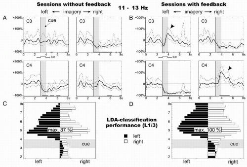
An example is shown in Figure 57.2 in the form of band power time courses of 11- to 13-Hz EEG activity. The ERD/ERS curves show different reactivity patterns during right and left motor imagery, displaying a significant band power decrease (ERD) over the contralateral hand area. It is of interest to note, first, that contralateral to the side of motor imagery an ERD and ipsilaterally an ERS were present and, second, that feedback enhanced the difference between both patterns, and therewith the classification accuracy (see also Ref. 50).
 Figure 57.2 Event-related desynchronization (ERD)/event-related synchronization (ERS) curves (11 to 13 Hz; 95% confidence intervals indicated) of one representative subject during imagined movements of the left versus right hand in sessions without feedback (A) and in sessions with continuously present feedback (C). Data were recorded from the sensorimotor cortex (C3, C4). The time period of cue presentation is indicated by a gray vertical bar. Examples of classification results of single trials (based on linear discriminant analysis, LDA) of two selected sessions: one without (B) and one with feedback (D). The x-axis denotes the average size of the distance function (resulting from LDA) for all left and right trials of one session (for details, see Ref. 69). In the session with feedback, the average distance corresponds to the average length of the feedback bar presented on the screen. Black bars indicate bar movements to the left side of the screen, white bars indicate bar movements to the right side. The y-axis displays the time points used for classification. The best classification accuracy for each session is indicated. |
The enhancement of oscillatory EEG activity (ERS) during motor imagery is a very important aspect in BCI research. For example, foot motor imagery can induce long-lasting beta oscillations during imagery (peri-imagery ERS; Fig. 57.3A) and/or short-lasting beta bursts after the end of the imagery process (postimagery ERS; Fig. 57.3B) over the foot representation area close to the vertex (29,51). The post-imagery ERS is dominant in the beta band with a maximum ˜2.5 seconds after brisk cue-paced imagery, can be detected with great accuracy (high rate of true positives, TP; see Fig. 57.3C) in the ongoing EEG and is therefore a good candidate to realize a one-channel EEG-based BCI (29,51).
Stay updated, free articles. Join our Telegram channel

Full access? Get Clinical Tree


