Epilepsy


Epilepsy


Carl W. Bazil

Shraddha Srinivasan

Timothy A. Pedley


INTRODUCTION

An epileptic seizure is the result of a temporary physiologic dysfunction of the brain caused by an abnormal, self-limited, and hypersynchronous electrical discharge of cortical neurons. There are many different kinds of seizures, each with characteristic behavioral changes and electrophysiologic disturbances that can usually be detected in scalp electroencephalography (EEG) recordings. The particular manifestations of any single seizure depend on several factors: whether most or only a part of the cerebral cortex is involved at the beginning, the functions of the cortical areas where the seizure originates, the subsequent pattern of spread of the electrical ictal discharge within the brain, and the extent to which subcortical and brain stem structures are engaged.

A seizure is a transient epileptic event, a symptom of disturbed brain function. Although seizures are the cardinal manifestation of epilepsy, not all seizures imply epilepsy. For example, seizures may be self-limited in that they occur only during the course of an acute medical or neurologic illness such as metabolic disarray or drug intoxication; they do not persist after the underlying disorder has resolved. Some people, for no discoverable reason, have a single unprovoked seizure. These kinds of seizures are not epilepsy.

Epilepsy is a chronic disorder, or group of chronic disorders, in which the indispensable feature is recurrence of seizures that are typically unprovoked and usually unpredictable. About 50 million people are affected worldwide. Each distinct form of epilepsy has its own natural history and response to treatment. This diversity presumably reflects the fact that epilepsy can arise from a variety of underlying conditions and pathophysiologic mechanisms, although most cases are classified as idiopathic (of presumed genetic origin) or cryptogenic (arising from a past injury that is not defined).


CLASSIFICATION OF SEIZURES AND EPILEPSY

Accurate classification of seizures and epilepsy is essential for understanding epileptic phenomena, developing a rational plan of investigation, making decisions about when and for how long to treat, choosing the appropriate antiepileptic drug (AED), and conducting scientific investigations that require delineation of clinical and EEG phenotypes.


CLASSIFICATION OF SEIZURES

The classification that is still followed today is the 1981 classification of epileptic seizures developed by the International League Against Epilepsy (ILAE; Table 58.1). This system classifies seizures by clinical symptoms supplemented by EEG data.


TABLE 58.1 International League Against Epilepsy Classification of Epileptic Seizures


I. Partial (focal) seizures


A. Simple partial seizures (consciousness not impaired)


1. With motor signs (including jacksonian, versive, and postural)


2. With sensory symptoms (including visual, somatosensory, auditory, olfactory, gustatory, and vertiginous)


3. With psychic symptoms (including dysphasia, dysmnesic, hallucinatory, and affective changes)


4. With autonomic symptoms (including epigastric sensation, pallor, flushing, pupillary changes)


B. Complex partial seizures (consciousness is impaired)


1. Simple partial onset followed by impaired consciousness


2. With impairment of consciousness at onset


3. With automatisms


C. Partial seizures evolving to secondarily generalized seizures


II. Generalized seizures of nonfocal origin (convulsive or nonconvulsive)


A. Absence seizures


1. With impaired consciousness only


2. With one or more of the following: atonic components, tonic components, automatisms, autonomic components


B. Myoclonic seizures


Myoclonic jerks (single or multiple)


C. Tonic-clonic seizures (may include clonic-tonic-clonic seizures)


D. Tonic seizures


E. Atonic seizures


III. Unclassified epileptic seizures


From the Commission on Classification and Terminology of the International League Against Epilepsy. Proposal for revised clinical and electroencephalographic classification of epileptic seizures. Epilepsia. 1981;22:489-501.


Inherent in the ILAE classification are two important physiologic principles. First, seizures are fundamentally of two types: those with onset limited to a part of one cerebral hemisphere (partial or focal seizures) and those that seem to involve the brain diffusely from the beginning (generalized seizures). Second, seizures are dynamic
and evolving; clinical expression is determined as much by the sequence of spread of electrical discharge within the brain as by the area where the ictal discharge originates. Variations in the seizure pattern exhibited by an individual imply variability in the extent and pattern of spread of the electrical discharge. Both generalized and partial seizures are further divided into subtypes. For partial seizures, the most important subdivision is based on consciousness, which is preserved in simple partial seizures or lost in complex partial seizures. Simple partial seizures may evolve into complex partial seizures, and either simple or complex partial seizures may evolve into secondarily generalized seizures. In adults, most generalized seizures have a focal onset whether or not this is apparent clinically. For generalized seizures, subdivisions are based mainly on the presence or absence and character of ictal motor manifestations.

The initial events of a seizure, described by either the patient or an observer, are usually the most reliable clinical indication to determine whether a seizure begins focally or is generalized from the moment of onset. Sometimes, however, a focal signature is lacking for several possible reasons:


  • The patient may be amnesic after the seizure, with no memory of early events.


  • Consciousness may be impaired so quickly or the seizure may generalize so rapidly that early distinguishing features are blurred or lost.


  • The seizure may originate in a brain region that is not associated with an obvious behavioral function. Thus, the seizure becomes clinically evident only when the discharge spreads beyond the ictal-onset zone or becomes generalized.

In 2010, the ILAE published a proposal for a revised classification of the epilepsies based on “terminology and concepts for organization.” As many epilepsy syndromes include both focal and generalized seizures, the classification distinguishing focal from generalized epilepsies was abandoned. In addition, a focal seizure was conceptualized as originating at some point within networks limited to one hemisphere. In contrast, a generalized seizure was conceptualized as originating at some point within, and then rapidly involving, networks distributed bilaterally. The three broad etiologic categories became genetic, structural/metabolic, and unknown, instead of cryptogenic, symptomatic, and idiopathic.


Partial Seizures

Simple partial seizures result when the ictal discharge occurs in a limited and often circumscribed area of cortex, the epileptogenic focus. Almost any symptom or phenomenon can be the subjective (aura) or observable manifestation of a simple partial seizure, varying from elementary motor (jacksonian seizures, adversive seizures) and unilateral sensory disturbance to complex emotional, psychoillusory, hallucinatory, or dysmnesic phenomena. Especially common auras include an epigastric rising sensation, fear, a feeling of unreality or detachment, déjà vu and jamais vu experiences, and olfactory hallucinations. Patients can interact normally with the environment during simple partial seizures except for limitations imposed by the seizure on specific localized brain functions.

Complex partial seizures, on the other hand, are defined by impaired consciousness and imply bilateral spread of the seizure discharge at least to basal forebrain and limbic areas. In addition to loss of consciousness, patients with complex partial seizures usually exhibit automatisms, such as lip smacking, repeated swallowing, clumsy perseveration of an ongoing motor task, or some other complex motor activity that is undirected and inappropriate. Postictally, patients are confused and disoriented for several minutes, and determining the transition from ictal to postictal state may be difficult without simultaneous EEG recording. Of complex partial seizures, 70% to 80% arise from the temporal lobe; foci in the frontal and occipital lobes account for most of the remainder.


Generalized Seizures

Generalized tonic-clonic (grand mal) seizures (Video 58.1) are characterized by abrupt loss of consciousness with bilateral tonic extension of the trunk and limbs (tonic phase), often accompanied by a loud vocalization as air is forcedly expelled across contracted vocal cords (epileptic cry), followed by synchronous muscle jerking ( clonic phase).

In some patients, a few clonic jerks precede the tonic-clonic sequence; in others, only a tonic or clonic phase is apparent. Postictally, patients are briefly unarousable and then lethargic and confused, often preferring to sleep. Many patients report inconsistent nonspecific premonitory symptoms (epileptic prodrome) for minutes to a few hours before a generalized tonic-clonic seizure. Common symptoms include illdefined anxiety, irritability, decreased concentration, and headache or other uncomfortable feelings; these are not auras.

Absence (petit mal) seizures are momentary lapses in awareness that are accompanied by motionless staring and arrest of any ongoing activity. Absence seizures begin and end abruptly; they occur without warning or postictal period. Mild myoclonic jerks of the eyelid or facial muscles, variable loss of muscle tone, and automatisms may accompany longer attacks. When the beginning and end of the seizure are less distinct, or if tonic and autonomic components are included, the term atypical absence seizure is used. Atypical absences are seen most often in children with epilepsy who are developmentally delayed or in epileptic encephalopathies, such as the Lennox-Gastaut syndrome (defined later in the chapter). Myoclonic seizures are characterized by rapid brief muscle jerks that can occur bilaterally, synchronously or asynchronously, or unilaterally. Myoclonic jerks range from isolated small movements of face, arm, or leg muscles to massive bilateral spasms simultaneously affecting the head, limbs, and trunk.

Atonic (astatic) seizures, (Video 58.2) also called drop attacks, are characterized by sudden loss of muscle tone, which may be fragmentary (e.g., head drop) or generalized, resulting in a fall. When atonic seizures are preceded by a brief myoclonic seizure or tonic spasm, an acceleratory force is added to the fall, thereby contributing to the high rate of self-injury with this type of seizure.


CLASSIFICATION OF EPILEPSY (EPILEPTIC SYNDROMES)

Attempting to classify the kind of epilepsy a patient has is often more important than describing seizures because the formulation includes other relevant clinical information of which the seizures are only a part. The other data include historical information (e.g., a personal history of brain injury or family history of first-degree relatives with seizures); findings on neurologic examination; and results of EEG, brain imaging, and biochemical studies.

The ILAE classification separates major groups of epilepsy first on the basis of whether seizures are partial (localization-related epilepsies) or generalized (generalized epilepsies) and second by cause ( idiopathic, symptomatic, or cryptogenic epilepsy). Subtypes of epilepsy are grouped according to the patient’s age and, in the case of localization-related epilepsies, by the anatomic location of the presumed ictal-onset zone.

Classification of the epilepsies has been less successful and more controversial than the classification of seizure types. A basic problem is that the classification scheme is empiric, with clinical and EEG data traditionally emphasized over anatomic, pathologic,
or specific etiologic information. This classification is useful for some reasonably well-defined syndromes, such as infantile spasms or benign partial childhood epilepsy with central-midtemporal spikes, especially because of the prognostic and treatment implications of these disorders. On the other hand, few epilepsies imply a specific disease or defect. A further drawback to the ILAE classification is that the same epileptic syndrome (e.g., infantile spasms or Lennox-Gastaut syndrome) may be “symptomatic” of a specific disease (e.g., tuberous sclerosis), considered “cryptogenic” on the basis of nonspecific imaging abnormalities, or categorized as “idiopathic.” Another biologic incongruity is the excessive detail in which some syndromes are identified, with specific entities culled from what are more likely simply different biologic expressions of the same abnormality (e.g., childhood and juvenile forms of absence epilepsy). As a result, a new classification of epilepsy syndromes has been proposed and is presently under discussion (Engel, 2006).

With the foregoing reservations, there is little question that defining common epilepsy syndromes has practical value. Table 58.2 gives a modified version of the current ILAE classification.


TABLE 58.2 Modified Classification of Epileptic Syndromes


I. Idiopathic epilepsy syndromes (focal or generalized)


A. Benign neonatal convulsions


1. Familial


2. Nonfamilial


B. Benign childhood epilepsy


1. With central-midtemporal spikes


2. With occipital spikes


C. Childhood/juvenile absence epilepsy


D. Juvenile myoclonic epilepsy (including generalized tonic-clonic seizures on awakening)


E. Idiopathic epilepsy, otherwise unspecified


II. Symptomatic epilepsy syndromes (focal or generalized)


A. West syndrome (infantile spasms)


B. Lennox-Gastaut syndrome


C. Early myoclonic encephalopathy


D. Epilepsia partialis continua


1. Rasmussen syndrome (encephalitic form)


2. Restricted form


E. Acquired epileptic aphasia (Landau-Kleffner syndrome)


F. Temporal lobe epilepsy


G. Frontal lobe epilepsy


H. Posttraumatic epilepsy


I. Other symptomatic epilepsy, focal or generalized, not specified


III. Other epilepsy syndromes of uncertain or mixed classification


A. Neonatal seizures


B. Febrile seizures


C. Reflex epilepsy


D. Other unspecified


SELECTED GENERALIZED EPILEPSY SYNDROMES


INFANTILE SPASMS (WEST SYNDROME)

The term infantile spasms denotes a unique age-specific form of generalized epilepsy that may be either idiopathic or symptomatic. When all clinical data are considered, including results of imaging studies, only about 15% of patients are now classified as idiopathic. Symptomatic cases result from diverse conditions, including cerebral dysgenesis, tuberous sclerosis, phenylketonuria, intrauterine infections, or hypoxic-ischemic injury.

Seizures are characterized by sudden flexor or extensor spasms that simultaneously involve the head, trunk, and limbs. The attacks usually begin before 6 months of age. The EEG is grossly abnormal, showing chaotic high-voltage slow activity with multifocal spikes, a pattern termed hypsarrhythmia. The treatment of choice is corticotropin or prednisone; spasms are notoriously refractory to conventional AEDs. Exceptions are topiramate and zonisamide, which have been shown to be an effective alternative to corticotropin in selected cases. Vigabatrin is also effective, especially in children with tuberous sclerosis. Patients on this drug must be monitored for visual field deficits. Although treatment with corticotropin, vigabatrin, zonisamide, or topiramate usually controls spasms and reverses the EEG abnormalities, it has little effect on long-term prognosis. Only about 5% to 10% of children with infantile spasms have normal or near-normal intelligence, and more than 66% have severe disabilities.


CHILDHOOD ABSENCE (PETIT MAL) EPILEPSY

This disorder begins most often between the ages of 4 and 12 years and is characterized predominantly by recurrent absence seizures, which, if untreated, can occur literally hundreds of times each day. EEG activity during an absence attack is characterized by stereotyped, bilateral, 3-Hz spike-wave discharges. Generalized tonic-clonic seizures also occur in 30% to 50% of cases. Most children are normal, both neurologically and intellectually. Ethosuximide and valproate are equally effective in treating absence seizures, but valproate or lamotrigine are preferable if generalized tonic-clonic seizures coexist. Topiramate, levetiracetam, and zonisamide may also be effective in generalized-onset seizures.


LENNOX-GASTAUT SYNDROME

This term is applied to a heterogeneous group of childhood epileptic encephalopathies that are characterized by mental retardation, uncontrolled seizures, and a distinctive EEG pattern. The syndrome is not a pathologic entity because clinical and EEG manifestations result from brain malformations, perinatal asphyxia, severe head injury, central nervous system (CNS) infection, or, rarely, a progressive degenerative or metabolic syndrome. A presumptive cause can be identified in 65% to 70% of affected children. Seizures usually begin before age 4 years, and about 25% of children have a history of infantile spasms. No treatment is consistently effective, and 80% of children continue to have seizures as adults. Best results are generally obtained with broad-spectrum AEDs, such as valproate, clobazam, lamotrigine, topiramate, or zonisamide. Rufinamide may be particularly effective for atonic seizures associated with this syndrome. Despite the higher incidence of severe side effects, fel bamate is often effective when these other agents do not result in optimal seizure control. Refractory cases may be considered for vagus nerve stimulation or anterior corpus callosotomy. Both of these are palliative procedures, and complete seizure control is rare.


JUVENILE MYOCLONIC EPILEPSY

The juvenile myoclonic epilepsy (JME) subtype of idiopathic generalized epilepsy most often begins in otherwise healthy individuals between the ages of 8 and 20 years. The fully developed syndrome comprises morning myoclonic jerks, generalized tonic-clonic seizures that occur just after waking, normal intelligence, a family history of similar seizures, and an EEG that shows generalized spikes, 4- to 6-Hz spike waves, and multiple spike (polyspike) discharges. The myoclonic jerks vary in intensity from bilateral massive spasms and falls to minor isolated muscle jerks that many patients consider nothing more than “morning clumsiness.” Linkage studies have produced conflicting results with different groups reporting susceptibility loci on chromosomes 6p, 5q, and 15q. A mutation in the α-1 subunit of the γ-aminobutyric acid (GABA)A receptor has been found in a large French Canadian family with JME but not in individuals with the common sporadic form of JME. Valproate is the treatment of choice and controls seizures and myoclonus in more than 80% of cases. Lamotrigine, zonisamide, levetiracetam, and topiramate can be equally effective in many patients, although lamotrigine sometimes exacerbates myoclonus.


SELECTED LOCALIZATION-RELATED EPILEPSY SYNDROMES


BENIGN FOCAL EPILEPSY OF CHILDHOOD

Several “benign” focal epilepsies occur in children, of which the most common is the syndrome associated with central-midtemporal spikes on EEG. This form of idiopathic focal epilepsy, also known as benign rolandic epilepsy, accounts for about 15% of all pediatric seizure disorders.

Onset is between 4 and 13 years of age; children are otherwise normal. Most children have attacks mainly or exclusively at night. Sleep promotes secondary generalization, so that parents report only generalized tonic-clonic seizures; any focal manifestations go unobserved. In contrast, seizures that occur during the day are clearly focal, with twitching of one side of the face; speech arrest; drooling from a corner of the mouth; and paresthesias of the tongue, lips, inner cheeks, and face. Seizures may progress to include clonic jerking or tonic posturing of the arm and leg on one side. Consciousness is usually preserved.

The interictal EEG abnormality is distinctive and shows stereotyped diphasic or triphasic sharp waves over the central-midtemporal (rolandic) regions. Discharges may be unilateral or bilateral. They increase in abundance during sleep and, when unilateral, switch from side to side on successive EEGs. In about 30% of cases, generalized spike-wave activity also occurs. The EEG pattern is inherited as an autosomal dominant trait with age-dependent penetrance. The inheritance pattern of the seizures, although clearly familial, is probably multifactorial and less well understood. More than half the children who show the characteristic EEG abnormality never have clinical attacks. Linkage has been reported in some families to chromosome 15q14.

The prognosis is uniformly good. Seizures disappear by mid to late adolescence in all cases. Seizures in many children appear to be self-limited, and not all children need AED treatment. Treatment can usually be deferred until after the second or third attack. Because seizures are easily controlled and self-limited, drugs with the fewest adverse effects, such as carbamazepine, oxcarbazepine, or gabapentin, should be used. Low doses, often producing subtherapeutic blood concentrations, are generally effective. Polytherapy should be avoided.


TEMPORAL LOBE EPILEPSY

This is the most common epilepsy syndrome of adults. In most cases, the epileptogenic region involves mesial temporal lobe structures, especially the hippocampus, amygdala, and parahippocampal gyrus. Seizures usually begin in late childhood or adolescence, and a history of febrile seizures is common. Virtually all patients have complex partial seizures, some of which secondarily generalize. Auras are frequent; visceral sensations are particularly common. Other typical behavioral features include a motionless stare, loss of awareness that may be gradual, and oral-alimentary automatisms, such as lip smacking (Video 58.3). A variable but often prolonged period of postictal confusion is the rule. Interictal EEGs show focal temporal slowing and epileptiform sharp waves or spikes over the anterior temporal region. AEDs are usually successful in suppressing secondarily generalized seizures, but most patients continue to have partial attacks. When seizures persist, anterior temporal lobe resection or selective amygdalohippocampectomy is the treatment of choice. For appropriate patients with mesial temporal sclerosis, ablation with laser or Gamma Knife has also been used, although it is not known whether these are as effective as more traditional resection in the long term. The results of a randomized controlled trial comparing seizure outcomes in patients treated either medically or surgically were striking: 58% of surgically treated patients were seizure free at 1 year compared with 8% of medically treated patients. Other series have shown that temporal lobe resection for refractory medial temporal lobe epilepsy associated with hippocampal sclerosis results in complete seizure control for at least 1 year in over 80% of patients. More controversial is the need for long-term anticonvulsant drug treatment after successful operation; a minority of patients may relapse several years later.


FRONTAL LOBE EPILEPSY

The particular pattern of the many types of frontal lobe seizures depends on the specific location where the seizure discharge originates and on the pathways subsequently involved in propagation. Despite this variability, the following features, when taken together, suggest frontal lobe epilepsy:


  • Brief seizures that begin and end abruptly with little, if any, postictal period


  • A tendency for seizures to cluster and to occur at night


  • Prominent, but often bizarre, motor manifestations, such as asynchronous thrashing or flailing of arms and legs; pedaling leg movements; pelvic thrusting; and loud, sometimes obscene, vocalizations, all of which may suggest psychogenic seizures (Video 58.4)


  • Minimal abnormality on scalp EEG recordings


  • A history of status epilepticus

Frontal lobe epilepsy occurs in some families as an autosomal dominant syndrome. In these patients, seizures almost always occur during sleep. Most patients respond well to medication.


POSTTRAUMATIC SEIZURES

Seizures occur within 1 year in about 7% of civilian and in about 34% of military head injuries. The differences relate mainly to the much higher proportion of penetrating wounds in military cases. The risk of developing posttraumatic epilepsy is directly related to the severity of the injury and also correlates with the total volume of brain lost as measured by computed tomography (CT). Depressed skull fractures may or may not be a risk; the rate of posttraumatic epilepsy was 17% in one series but not increased above control
levels in another. Head injuries are classified as severe if they result in brain contusion, intracerebral or intracranial hematoma, unconsciousness, or amnesia for more than 24 hours or in persistent neurologic abnormalities, such as aphasia, hemiparesis, or dementia. Mild head injury (brief loss of consciousness, no skull fracture, no focal neurologic signs, no contusion or hematoma) does not increase the risk of seizures significantly above general population rates.

Nearly 60% of those who have seizures have the first attack in the first year after the injury. In the Vietnam Head Injury Study, however, more than 15% of patients did not have epilepsy until 5 or more years later. Posttraumatic seizures are classified as early (within the first 1 to 2 weeks after injury) or late. Only recurrent late seizures (those that occur after the patient has recovered from the acute effects of the injury) should be considered posttraumatic epilepsy. Early seizures, however, even if isolated, increase the chance of developing posttraumatic epilepsy. About 70% of patients have partial or secondarily generalized seizures. Impact seizures occur at the time of or immediately after the injury. These attacks are attributed to an acute reaction of the brain to trauma and do not increase the risk of later epilepsy.

Overt seizures should be treated according to principles reviewed later in this chapter. The most controversial issue concerns the prophylactic use of AEDs to retard or abort the development of subsequent seizures. Based on the data of Temkin et al. (1990), we recommend treating patients with severe head trauma, as just defined, with an antiepileptic medication for the first week after injury to minimize complications from seizures occurring during acute management. If seizures have not occurred, we do not continue medication beyond the initial 1 to 2 weeks because evidence does not show that longer treatment prevents the development of later seizures or of posttraumatic epilepsy. Data suggest that valproate is less effective than phenytoin in suppressing acute seizures and is also ineffective in preventing the development of posttraumatic epilepsy.


EPILEPSIA PARTIALIS CONTINUA

Epilepsia partialis continua (EPC) refers to unremitting motor seizures involving part or all of one side of the body. They typically consist of repeated clonic or myoclonic jerks that may remain focal or regional or may march from one muscle group to another, with the extent of motor involvement waxing and waning in endless variation (Video 58.5). In adults, EPC occurs in diverse settings, such as with subacute or chronic inflammatory diseases of the brain (Kozhevnikov Russian spring-summer encephalitis, Behçet disease) or with acute strokes, metastases, and metabolic encephalopathies, especially hyperosmolar nonketotic hyperglycemia.

The most distinctive form of EPC, known as the Rasmussen syndrome, occurs in children; it usually begins before the age of 10 years. The underlying disorder is chronic focal encephalitis, although an infectious agent has not been identified consistently. About two-thirds of patients report an infectious or inflammatory illness 1 to 6 months before onset of EPC. Generalized tonic-clonic seizures are often the first sign and appear before the EPC establishes itself. About 20% of cases begin with an episode of convulsive status epilepticus. Slow neurologic deterioration inevitably follows, with development of hemiparesis, mental impairment, and, usually, hemianopia. If the dominant hemisphere is affected, aphasia occurs. EEGs are always abnormal, but findings are not specific, and they frequently do not correlate with clinical manifestations. Magnetic resonance imaging (MRI) may be normal early but later shows unilateral cortical atrophy and signal changes consistent with gliosis. Autoantibodies to the GluR3 protein of the glutamate receptor have been found in some patients, suggesting that autoimmunity may play a role in the pathogenesis of the disorder in some patients, and immunotherapy is sometimes beneficial. AEDs are usually ineffective in controlling seizures and preventing progression of the disease, as are corticosteroids and antiviral agents. When seizures have not spontaneously remitted by the time there is a severe degree of hemiparesis, functional hemispherectomy can control seizures and leads to substantial intellectual improvement in many patients. Controversy arises in the decision about the best time for hemispherectomy, for example, whether it should be performed earlier before there is serious motor or language impairment.


EPIDEMIOLOGY

In the United States, about 6.5 persons per 1,000 population are affected with recurrent unprovoked seizures, so-called active epilepsy. Based on 1990 census figures, age-adjusted annual incidence rates for epilepsy range from 31 to 57 per 100,000 in the United States (Fig. 58.1). Incidence rates are highest among young children and the elderly; epilepsy affects males 1.1 to 1.5 times more often than females.

Complex partial seizures are the most common seizure type among newly diagnosed cases, but age-related variability occurs in the proportions of different seizure types (Fig. 58.2). The cause of epilepsy also varies somewhat with age. Despite advances in diagnostic capabilities, however, the “unknown” etiologic category remains larger than any other for all age groups (Fig. 58.3). Cerebrovascular disease, associated developmental neurologic disorders (e.g., cerebral palsy and mental retardation), and head trauma are the other most commonly identified causes.

Although defined genetic disorders account for only about 1% of epilepsy cases, heritable factors are important. Monozygotic twins have a much higher concordance rate for epilepsy than do dizygotic twins. By age 25 years, nearly 9% of children of mothers with epilepsy and 2.4% of children of affected fathers develop epilepsy. The reason for an increased risk of seizures in children of women with epilepsy is not known.

Some forms of epilepsy are more heritable than others. For example, children of parents with absence seizures have a higher risk of developing epilepsy (9%) than do offspring of parents with other types of generalized seizures or partial seizures (5%). As a general rule, though, even offspring born to a high-risk parent have a 90% or greater chance of being unaffected by epilepsy.


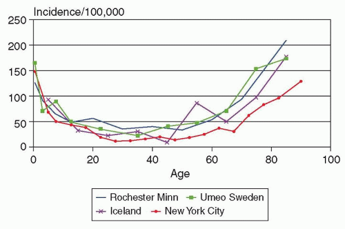
FIGURE 58.1 Age-specific incidence of epilepsy in Rochester, Minnesota, 1935-1984. (From Hauser WA, Annegers JF, Kurland LT. Incidence of epilepsy and unprovoked seizures in Rochester, Minnesota: 1935-1984. Epilepsia. 1993;34(3): 453-468.)


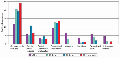
FIGURE 58.2 Proportion of seizure types in newly diagnosed cases of epilepsy in Rochester, Minnesota, 1935-1984. (From Hauser WA, Annegers JF, Kurland LT. Incidence of epilepsy and unprovoked seizures in Rochester, Minnesota: 1935-1984. Epilepsia. 1993;34(3):453-468.)

Many persons who experience a first unprovoked seizure never have a second. By definition, these people do not have epilepsy and generally do not require long-term drug treatment. Unfortunately, our ability to identify such individuals with accuracy is imperfect. Treatment decisions must be based on epidemiologic and individual considerations. Some seizure types, such as absence and myoclonic, are virtually always recurrent by the time the patient is seen by a physician. On the other hand, patients with convulsive seizures may seek medical attention after a first occurrence because of the dramatic nature of the attack. Prospective studies of recurrence after a first seizure indicate a 2-year recurrence risk of about 40%, which is similar in children and adults. The risk is lowest in people with an idiopathic generalized first seizure and normal EEG (about 24%), higher with idiopathic generalized seizures and an abnormal EEG (about 48%), and highest with symptomatic (i.e., known preceding brain injury or neurologic syndrome) seizures and an abnormal EEG (about 65%). Epileptiform, but not nonepileptiform, EEG abnormalities impart a greater risk for recurrence. If the first seizure is a partial seizure, the relative risk of recurrence is also increased. The risk for further recurrence after a second unprovoked seizure is greater than 80%; a second unprovoked seizure is, therefore, a reliable marker of epilepsy.


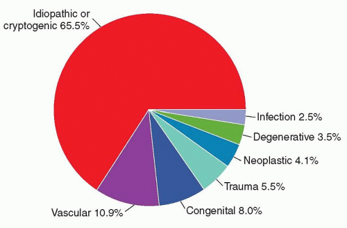
FIGURE 58.3 Etiology of epilepsy in all cases of newly diagnosed seizures in Rochester, Minnesota, 1935-1984. (From Hauser WA, Annegers JF, Kurland LT. Incidence of epilepsy and unprovoked seizures in Rochester, Minnesota: 1935-1984. Epilepsia. 1993;34(3):453-468.)

About 4% of persons living to age 74 years have at least one unprovoked seizure. When provoked seizures (i.e., febrile seizures or those related to an acute illness) are included, the likelihood of experiencing a seizure by age 74 years increases to at least 9%. The risk of developing epilepsy is about 3% by age 74 years.

Of persons with epilepsy, 60% to 70% achieve remission of seizures with AED therapy. Factors that favor remission include an idiopathic (or cryptogenic) form of epilepsy, normal findings on neurologic examination, and onset in early to middle childhood (except neonatal seizures). Unfavorable prognostic factors include partial seizures, an abnormal EEG, and associated mental retardation or cerebral palsy (Table 58.3). Those who fail to achieve remission have drug-resistant epilepsy and may be candidates for alternative therapies, including surgery or devices as well as additional drug treatment. Drug-resistant epilepsy is defined by the ILAE as “failure of adequate trials of two tolerated and appropriately used antiepileptic drugs to achieve sustained seizure freedom.”


TABLE 58.3 Predictors of Intractable Epilepsy


Very young at onset (<2 yr)


Frequent generalized seizures


Failure to achieve control readily


Evidence of brain damage


A specific cause of the seizures


Severe EEG abnormality


Low IQ


Atonic atypical absence seizures


EEG, electroencephalogram.


Mortality is increased in persons with epilepsy, but the risk is incurred mainly by symptomatic cases in which higher death rates are related primarily to the underlying disease rather than to epilepsy. Accidental deaths, especially drowning, are more common, however, in all patients with epilepsy. Sudden unexplained death is nearly 25 times more common in patients with epilepsy than in the general population; estimates of incidence rates range from 1 in 500 to 1 in 2,000 per year. Severe epilepsy, uncontrolled generalized convulsions, especially nocturnal, and need for multiple AEDs are risk factors.


INITIAL DIAGNOSTIC EVALUATION

The diagnostic evaluation has three objectives: to determine if the patient has epilepsy; to classify the type of epilepsy and identify an epilepsy syndrome, if possible; and to define the specific underlying cause. Accurate diagnosis leads directly to proper treatment and formulation of a rational plan of management. The differential diagnosis is considered in Section II, Chapter 5.

Because epilepsy comprises a group of conditions and is not a single homogeneous disorder and because seizures may be symptoms of both diverse brain disorders and an otherwise normal brain, it is neither possible nor desirable to develop inflexible guidelines for what constitutes a standard or minimal diagnostic evaluation. The clinical data from the history and physical examination should allow a reasonable determination of probable diagnosis, seizure and epilepsy classification, and likelihood of underlying brain disorder. Based on these considerations, diagnostic testing should be undertaken selectively.


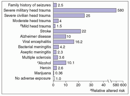
FIGURE 58.4 Risk factors for epilepsy. aRelative to people without these adverse exposures. bNot statistically significant. cOne pint of 80 proof, 2.5 bottles of wine. (From Hauser WA, Hesdorffer DC. Epilepsy: Frequency, Causes and Consequences. New York: Demos; 1990.)


ELECTROENCEPHALOGRAPHY

Because epilepsy is fundamentally a physiologic disturbance of brain function, the EEG is the most important laboratory test in evaluating patients with seizures. The EEG helps both to establish the diagnosis of epilepsy and to characterize specific epileptic syndromes. EEG findings may also help in management and in prognosis.

Epileptiform discharges (spikes and sharp waves) are highly correlated with seizure susceptibility and can be recorded on the first EEG in about 50% of patients. Similar findings are recorded in only 1% to 2% of normal adults and in a somewhat higher percentage of normal children. When multiple EEGs are obtained, epileptiform abnormalities eventually appear in 60% to 90% of adults with epilepsy, but the yield of positive studies does not increase substantially after three or four tests. Prolonged ambulatory or inpatient recordings increase the yield of interictal epileptiform abnormalities both because of the longer sampling times but also because complete sleep-wake cycles are included. It is important to remember, therefore, that 10% to 40% of patients with epilepsy do not show epileptiform abnormalities on routine EEG. Thus, a normal or nonspecifically abnormal EEG never excludes the diagnosis. Sleep, hyperventilation, photic stimulation, and special electrode placements are routinely used to increase the probability of recording epileptiform abnormalities. Different and distinctive patterns of epileptiform discharge occur in specific epilepsy syndromes as summarized in Chapter 25.


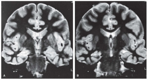
FIGURE 58.5 Mesial temporal sclerosis. A,B: Short-tau inversion recovery (STIR) coronal magnetic resonance images through the temporal lobes show increased signal and decreased size of right hippocampus as compared with left. These findings are characteristic of mesial temporal sclerosis. Note incidental focal dilatation of left choroid fissure, which represents a choroid fissure cyst and is a normal variant. (Courtesy of Dr. S. Chan, Columbia University College of Physicians and Surgeons, New York, NY.)


BRAIN IMAGING

MRI should be performed in all patients older than age 18 years and in children with abnormal development, abnormal findings on physical examination, or seizure types that are likely to be manifestations of symptomatic epilepsy. CT will often miss common epileptogenic lesions such as hippocampal sclerosis, cortical dysplasia, and cavernous malformations. Because CT is very sensitive for detecting brain calcifications, a noncontrast CT (in addition to MRI) may be helpful in patients at risk for neurocysticercosis.

Routine imaging is not necessary for children with idiopathic epilepsy, including the benign focal epilepsy syndromes (see section “Benign Epilepsy Syndromes”). Brain MRI, although more costly, is more sensitive than CT in detecting potentially epileptogenic lesions, such as cortical dysplasia, hamartomas, differentiated glial tumors, and cavernous malformations. Both axial and coronal planes should be imaged using both T1 and T2 sequences. Gadolinium injection does not increase the sensitivity for detecting cerebral lesions but may assist in differentiating possible causes.

Imaging in the coronal plane perpendicular to the long axis of the hippocampus and other variations in technique have improved the detection of hippocampal atrophy and gliosis, findings that are highly correlated with mesial temporal sclerosis (Fig. 58.5) and an epileptogenic temporal lobe. An even more sensitive measure of hippocampal atrophy is MRI measurement of the volume of the hippocampus. Hippocampal volume measurements in an individual patient then can be compared with those of normal control subjects. In patients being considered for surgery, interictal
positron emission tomography (PET) scans can add valuable localizing information, especially when the MRI scan is negative. Singlephoton emission computed tomography (SPECT) scans are also used, although resolution is less than either MRI or PET. Subtraction ictal SPECT co-registered with MRI (SISCOM) is also helpful in localizing the epileptogenic brain region in some cases.


OTHER LABORATORY TESTS

Routine blood tests are necessary in newborns and in older patients with acute or chronic systemic disease to detect abnormal electrolyte, glucose, calcium, or magnesium values or impaired liver or kidney function that may contribute to seizure occurrence. They are rarely diagnostically useful in healthy children or adults. Serum electrolytes, liver function tests, and a complete blood count (CBC) should be obtained when infectious or metabolic abnormalities are suspected, but they are useful mainly as baseline studies before initiating AED treatment.

Any suspicion of meningitis or encephalitis mandates lumbar puncture. Urine or blood toxicologic screens should be considered when otherwise unexplained new-onset generalized seizures occur.


Jul 27, 2016 | Posted by in NEUROLOGY | Comments Off on Epilepsy

Full access? Get Clinical Tree

Get Clinical Tree app for offline access