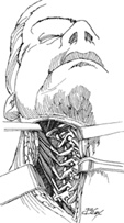
17 Daniel K. Resnick The vertebral artery runs through the foramen transversarium of the first six vertebral bodies, loops around the superolateral margin of the lamina of C1, and then enters the skull through the foramen magnum. The vertebral artery is vulnerable to injury following facet subluxation injuries, as well as during anterior cervical diskectomy (and anterior microforaminotomy), lateral mass and C3–C6 pedicle screw placement, and exposure and fixation of the craniocervical junction. Occasionally, exposure of the vertebral artery may be required for direct repair or occlusion following iatrogenic injury. Appreciation of the anatomy of the vertebral artery and the surrounding bony structures is essential for complication avoidance. Anatomic variation is most commonly seen at the level of C2, where 15 to 20% of patients may have aberrant vessels that preclude pars interarticularis or pedicle screw placement. Preoperative study and knowledge of alternative fixation techniques decrease the incidence of vertebral artery injury. The most important principle in managing a vertebral artery injury is to avoid a bilateral injury at all costs. Management of vertebral artery injuries requires immediate control of hemorrhage, the use of alternative fixation techniques, prompt assessment of cerebrovascular competence, and if necessary repair or occlusion of the vertebral artery. Iatrogenic injury of the vertebral artery. Once hemostasis is ensured, further efforts to explore the vertebral artery are reserved for cases with demonstrated or suspected neurologic compromise. The loss of a single vertebral artery is usually well tolerated. The loss of both vertebral arteries is usually fatal. Embolism and pseudoaneurysm formation are late complications of injury that may require arterial reconstruction, bypass, or occlusion. In the case of iatrogenic injury, the patient is positioned for the index procedure. If elective exploration is performed, the patient may be positioned supine with the head turned away from the operative site. Close monitoring of blood pressure to preserve cerebral perfusion pressure is mandatory. Complication avoidance is much preferred to complication management. Careful preoperative assessment of the location of the vertebral artery in relationship to anticipated sites of operative manipulation is essential. If a vertebral artery injury is identified or suspected, the contralateral vertebral artery must be preserved. This may result in unilateral fixation (side of the injury) or the need for external immobilization. Immediate postoperative assessment of cerebrovascular anatomy and reserve should be coordinated with an experienced cerebrovascular neurosurgeon. Blood loss may be rapid and significant. Depending on the site of injury, bleeding may be controlled with tamponade, packing with hemostatic agents, or simply by screw placement. Local exploration and attempts at repair may be possible in rare instances. Such attempts should not be undertaken without prior cerebrovascular experience and an adequately prepared operating room staff. Endovascular techniques for occlusion or stenting of the artery should be considered as valuable treatment options. If exploration of the artery is undertaken, the surgeon must be prepared to deal with the risk of significant bleeding, hemodynamic lability, and the risk of embolization. Injuries to the vertebral artery at or above C2 usually are encountered during the initial exposure (injuring the vessel along the rostral border of the C1 lamina) or by inadvertent laceration of the artery by a drill, tap, or screw. If the vessel is injured during the initial exposure, the site of injury is obvious and direct tamponade followed by local dissection will expose the area of injury. If the bleeding is through a hole in the bone, screw placement will generally achieve hemostasis. If the injury occurs during a posterior fixation procedure below C2, injury is likely related to screw placement. Hemostasis should be obtained with screw placement; the ipsilateral construct is rapidly completed, and the contralateral vertebral artery is preserved. Postoperative imaging is obtained. Conventional angiographic study is preferred, as the visualization of the artery is unaffected by metallic artifact, and therapeutic maneuvers such as stenting or occlusion may be performed contemporaneously. If the artery is occluded, anticoagulation or bypass should be considered in cooperation with a cerebrovascular specialist. In the asymptomatic patient, no therapy may be required. Revascularization options may also include endovascular techniques and bypass procedures such as the occipital artery-posterior inferior cerebellar artery bypass. If an injury occurs during an anterior procedure, such as during an anterior cervical diskectomy or an anterior microforaminotomy, then exploration may be feasible. Exploration may be considered when hemostasis cannot be adequately obtained locally, or when immediate preservation of the artery is deemed to be essential (dominant vertebral artery, known contralateral occlusion) and the surgeon has some experience with vascular repair. The vertebral artery may be exposed over several segments from an anterolateral approach. It may be accomplished through an extension of the standard anterior cervical approach. The incision is made along the border of the sternocleidomastoid, and may be extended to just posterior to the mastoid process. The precervical fascia is divided and the plane medial to the sternocleidomastoid is exploited to expose the prevertebral fascia overlying the spine. The longus colli is mobilized to expose the transverse processes (Fig. 17.1) and the transverse foramina are opened using a drill or Kerrison rongeurs. For more rostral exposure of the vertebral artery, a more lateral approach is preferred (Fig. 17.2). The sternocleidomastoid muscle is detached from the mastoid process and reflected caudally (leave a cuff of muscle attached to the mastoid for later closure). Identification and preservation of the spinal accessory nerve will avoid a postoperative shoulder drop (Fig. 17.3). As the sternocleidomastoid muscle is retracted, the transverse processes of the upper three or four vertebral bodies will be palpable, as will the pulsatile vertebral artery. Serial section of the splenius capitis and levator scapula muscles will expose the lateral aspect of the vertebral artery as it runs in the transverse foramen and then loops across C1 (Fig. 17.4).
Exposure of the Vertebral Artery
Description
Key Principles
Expectations
Indications
Contraindications
Special Considerations
Special Instructions, Positioning, and Anesthesia
Tips, Pearls, and Lessons Learned
Difficulties Encountered
Key Procedural Steps
Bailout, Rescue, Salvage Procedures

Stay updated, free articles. Join our Telegram channel

Full access? Get Clinical Tree


