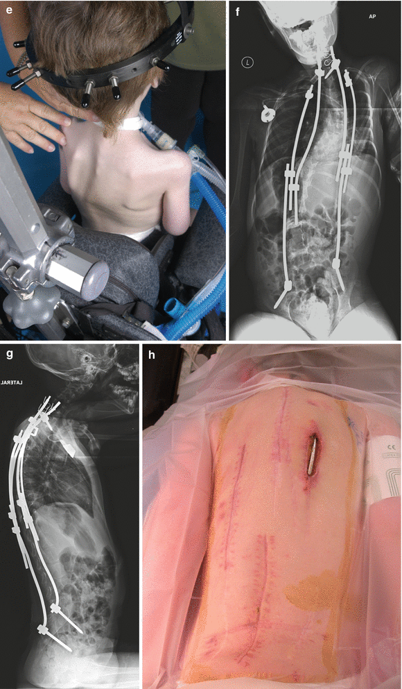
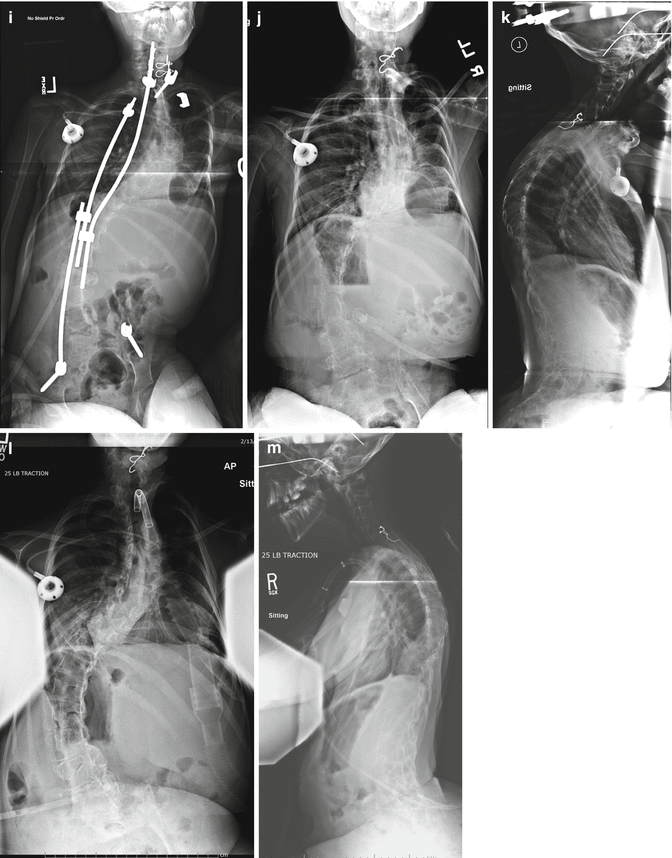
Fig. 30.1
(a, b) A 4-year-old boy with collapsing kyphoscoliosis due to congenital myopathy (March 2007); (c, d) scoliosis curves measuring 95 and 90°, with 85° kyphosis; (e) correction after 2 months HGT; (f, g) placement of rib-pelvis and spine-pelvis construct (August 2007), followed by one interval lengthening; (h) rod erosion through skin (December 2008). Treatment by wound vac: (i) removal of right rods (January 2009); (j, k) removal of all implants (January 2010) due to unresolving wound sepsis. HGT was re-started: (l, m) 4 years later (January 2014). Continuous traction has stabilized deformity
30.2 Indications
Patients with early onset spinal deformity often present with co-morbidities and physical characteristics which can significantly challenge and compromise any surgical treatment plan. For example, those with syndromic or “exotic” diagnoses possess diminutive osteopenic bony elements, which can severely limit acute deformity correction due to weakness of the bone-implant interface. Frequently, their deformity is rigid and/or kyphotic, in which case posterior distractive methods of correction (rod systems, VEPTR’s) are compromised by the need for extreme contouring of the expandable device, leading to ineffective distraction and proximal anchor failure by posterior cut-out, if not peri-operatively, then later by fatigue “plowing” or fracture due to the unfavorable biomechanical forces – especially in kyphosis (Fig. 30.2).
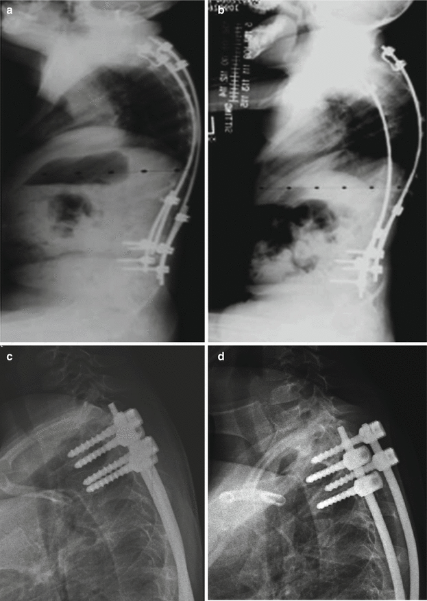

Fig. 30.2
(a, b) Proximal rib hook anchors failing over a period of 1 year in a collapsing kyphotic deformity; (c, d) thoracic pedicle screw pullout within the first year after implantation in an ambulatory patient
Neurological injury from acute correction is always a concern, especially if severe deformity requires canal manipulation by osteotomy or vertebral resection to achieve it. Relative canal stenosis is another source of neurological risk with acute correction, especially if previous fusion produces cord compression at a junctional segment due to fusion mass overgrowth or a juxta-fusion hypermobile segment [1, 2].
Patients with severe thoracic deformity, especially as a sequela of neglect or ineffective previous treatment, may present with respiratory impairment, in which case HGT is indicated as a preliminary step to improve respiratory mechanics and make them a more suitable surgical candidate. Such respiratory compromise, as well as hypotonia and weakness, chest wall defects, skin intolerance or anesthesia, and mental retardation may eliminate external means of deformity control, such as bracing or casting, from consideration.
Patients with severe deformity are also not candidates for bracing or casting purely from biomechanical considerations related to curve magnitude. Curves exceeding approximately 53° are corrected more effectively by longitudinal distraction forces [3], rather than laterally applied transverse forces realized with a cast or brace. Large, stiff curves thus may not benefit from use of the latter, being poorly tolerated due to excess skin pressure associated with inefficient transverse loads, as well as rib and chest wall deformation caused by the lateral rib pressure. In these instances, HGT is an effective method to achieve deformity correction, and indirectly, improve respiratory mechanics [4, 5]. We have noted up to 10 % increase in predicted vital capacity acutely in several patients who benefit from elongation of the chest wall associated with the spinal elongation/correction (Fig. 30.3). Improving the restrictive component of the deformity probably results from more efficient diaphragmatic excursion in the elongated trunk as well as from rib separation on the concavity, providing more effective inspiration and consequently respiration. This appears to be the physiologic explanation for this acute vital capacity increase during HGT.
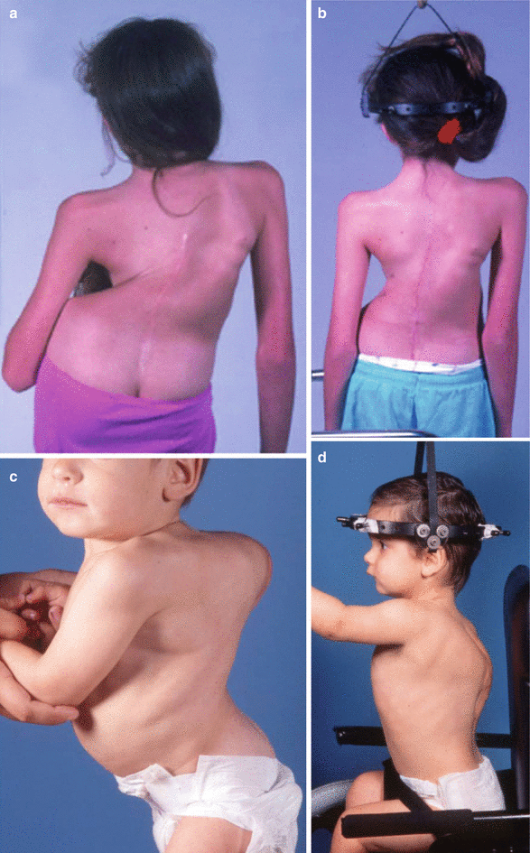

Fig. 30.3
(a, b) Elongation of the thorax by HGT following osteotomies of previous fusion mass; (c, d) elongation with kyphosis correction
30.3 Contraindications
HGT has been found to be almost uniformly safe [4–7]. The only absolute contraindications to its use would be bone stock in the skull insufficient to gain halo purchase due to underlying diagnoses such as osteogenesis imperfecta or fibrous dysplasia (Fig. 30.4); presence of an intra- or extra-medullary lesion (tumor, syrinx), with or without pre-existing neurologic deficit (Fig. 30.5); severe canal distortion with stenosis [1]. Otherwise, any patient with severe rigid deformity with or without kyphosis, potential or actual thoracic insufficiency syndrome, osteopenia, and increased potential neurological risk from acute instrumented correction is a candidate for HGT as a preliminary step before other operative treatment, to reduce the occurrence of instrumentation or neurological complications and to improve respiratory function and suitability for general anesthesia.
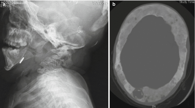
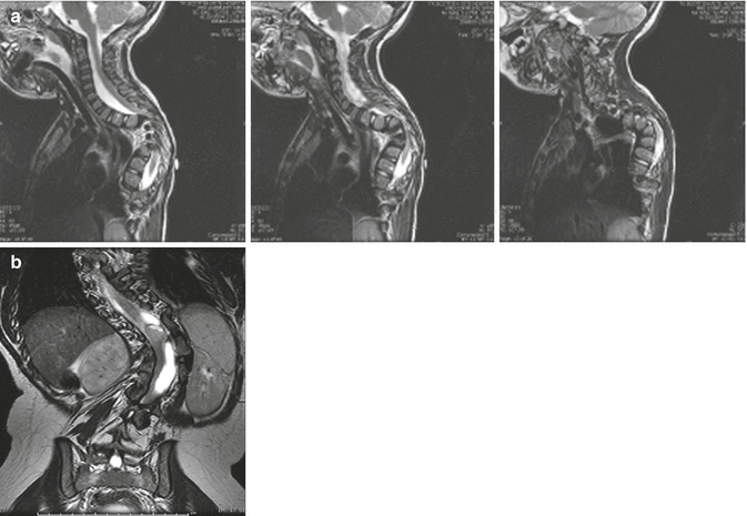

Fig. 30.4
(a) Absence of occipital bone in a patient with Loeys-Dietz syndrome. This patient is unsuitable for halo placement; (b) severe skull involvement with fibrous dysplasia, also unsuitable for halo placement

Fig. 30.5
(a) Cord compression with paraparesis in a 6-year-old patient with Pierre-Robin syndrome. Surgical decompression, not HGT, is indicated; (b) MRI revealing cystic astrocytoma in a 7-year-old male who was neurologically normal. HGT was started and he rapidly became paraparetic, which did not resolve with discontinuing the traction
30.4 Technique
HGT is not a new method of treatment of spinal deformity, having been developed soon after the halo apparatus was first described in the 1960s at Rancho Los Amigos Hospital [8]. Stagnara [9] popularized the gravity-traction method, which was introduced at our institution after it was demonstrated to the author by Klaus Zielke in 1984 during a visit to the latter’s clinic in Germany. The indications for which these early authors used HGT were essentially the same as its use for us today – neuromuscular “collapsing” deformity in the Rancho experience and as an adjunct to neglected deformity in older patients with respiratory insufficiency, as well as in the young patient with syndromic or exotic spinal deformity.
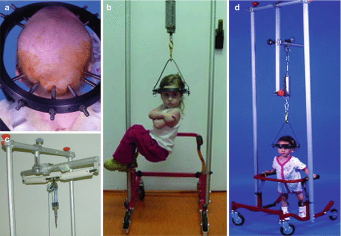
Halo application requires general anesthesia for children of this age group, using the maximum number of pins possible [5, 10] (Fig. 30.6a). Experience has shown that the use of numerous pins actually decreases the incidence of pin infection or loosening of any single pin. Pin direction should also be as perpendicular to the skull as possible [11, 12]. Pins are tightened to a torque approximately equaling the age of the child up to a maximum of 8 inch-pounds, e.g., a 4-year-old patient’s pins are tightened to 4 inch-pounds of torque, using a calibrated torque wrench. Because of variation in skull thickness and location of sutures, computed tomography (CT) scanning of the skull has been recommended to control pin placement [13, 14], but in practice such thickness determination has not altered intended pin location when multiple pins are used and the penetration is controlled by torque determination. Frontal and occipital areas are commonly adequate for safe, secure placement. Upright overhead traction via a traction bale attached to a wheelchair or standing frame, using a spring-loaded fish scale or other dynamic traction device (Fig. 30.6b–d) is begun the following day, initially with 5–10 pounds of traction. The amount of time and weight is increased to tolerance under careful neurological surveillance. All patients should have cranial nerve testing once a shift while upright in traction, as well as motor and sensory testing of upper and lower extremities, especially during the phase of increasing traction. Eventually traction force exceeding 50 % of body weight may be achieved, with cervical pain being the usual limiting factor. The goal of just lifting the patient’s buttocks off the wheelchair seat while sitting, or being on tiptoes in the standing frame can usually be attained within 2 weeks (see Fig. 30.6d). The use of nighttime traction, making the treatment program more or less continuous, can be added by providing a cervical traction frame to the patient’s bed, usually a gatched bed with the head portion elevated to act as a counter-traction [15]. Out-patient (home) traction can be attempted if caregivers are appropriately trained and vigilant. Radiographs are obtained every 3–4 weeks until a plateau of correction is reached. Remaining in traction without complication and with gradual deformity improvement over a period of 6 months is not unusual. In selected instances, in patients deemed too fragile for operative treatment or in whom operative treatment has failed and been abandoned, we have successfully treated severe uncorrectable deformity by extending HGT treatment indefinitely (Fig. 30.1).
