Fig. 31.1
Black: osseous part of the vertebrae. Dark grey: growing structures Light grey: disc structures. (a) The HV to be removed. (b) The surrounding growing structures of the HV remain after resection. (c) Compression force is applied. (d) The gap is spontaneously filled by new bone formation
Whether the approach is single posterior or anterior and posterior and whether hooks or screws are used, correction is achieved through compression forces on the convexity. At the end of the procedure, the surrounding growth structure of the HV is still active.
The empty space will be filled with spontaneous new bone formation.
The positive consequence is the stability of the vertebral column in its anterior aspect, but a possible negative consequence is that there may be “recurrence,” at least partial of the HV. That explains the observed “new bone mass at the site of the HV” as reported by Ruff and Harms [38].
This option is the one used in the so called “eggshell procedure” and is usually performed by a posterior-only approach [32].
Second Option Is to Remove Both the Osseous Hemivertebral Body and the Surrounding Growth Structures of the HV (Fig. 31.2)
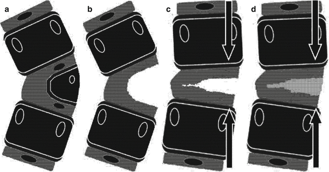
Fig. 31.2
Black: osseous part of the vertebrae. Dark grey: growing structures. Light grey: disc structures. (a) The HV to be removed. (b) The surrounding growing structures of the HV are removed. (c) Compression force is applied. (d) The gap is spontaneously filled by a fibrous scar (Courtesy of Gerard Bollini)
Whether the approach is single posterior or anterior and posterior and whether hooks or screws are used again, correction is achieved through compression forces on the convexity. At the end of the procedure, the growth structure of the adjacent vertebrae is still active.
The negative consequence of such a procedure is that the gap created anteriorly is filled with a fibrous scar whose mechanical stability is doubtful.
The relative positive consequence is that growth may occur between the resting endplates of the vertebrae adjacent to the HV.
The term “relative” is used because such a growth is correlated with distraction forces on the anterior aspect of the spinal column which could turn on posterior hardware breakage or dislodgement.
It is impossible to get an anterior convex arthrodesis between the two vertebrae adjacent to the HV and in the meantime to observe further growth coming from these same two vertebrae as stated by Ruf (Fig 31.3).
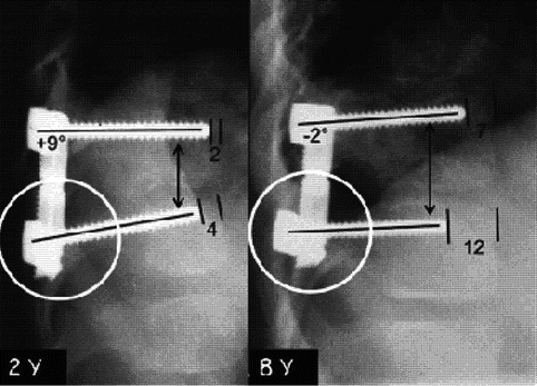

Fig. 31.3
Postoperative radiograph after HV resection, at 2 years of age on the left and at 6 years follow-up on the right. Although an arthrodesis has been performed posteriorly, there is still growth from the two adjacent vertebral bodies anteriorly (note the dislodgement of the bolt of the lower screw in the white circles) (Reprinted from Ruf and Harms [40]. With permission from Wolters Kluwer Health)
Third Option Is to Remove Both the Whole HV (Osseous Hemivertebral Body + Surrounding Growth Structures) and the Endplates of the Adjacent Vertebrae on the Convex Side of the Scoliosis (Fig. 31.4)
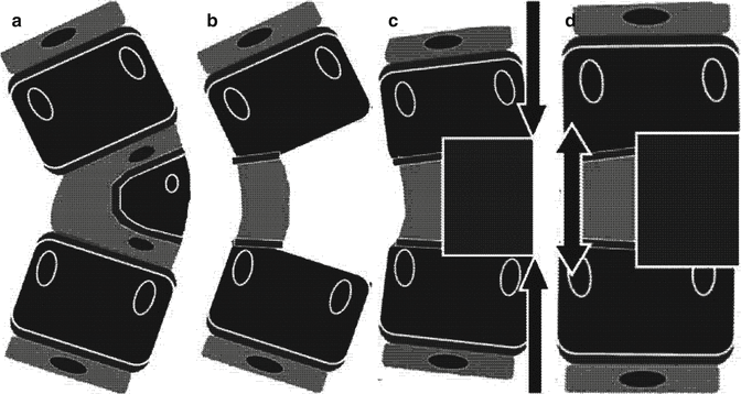
Fig. 31.4
Black: osseous part of the vertebrae. Dark grey: growing structures. Light grey: disc structures. (a) The HV to be removed. (b) The surrounding growing structures of the HV are removed as well as the disc and the convex growing structures of the adjacent vertebrae. (c) Compression force is applied. (d) The gap is filled by a bone graft (Courtesy of Gerard Bollini)
It is the only option which allows a true convex epiphysiodesis under the condition that bone grafting is performed between the two adjacent vertebral bodies on its convex aspect.
From these three options, whatever the option chosen, the main point to understand is that at the end of such procedures and after having applied compression forces on the posterior aspect of the vertebral column, there is still a gap between the two adjacent vertebral bodies.
The gap can be filled with a bone graft (Option 3) if an anterior and a posterior approach are performed in the same session and in such case hooks or screws can be indifferently used.
But in a posterior-only approach, screws rather than hooks are used to avoid a secondary kyphotic deformity and/or a pseudoarthrosis. Hooks and rods do not provide significant enough stabilization as shown in Figs. 31.5 and 31.6, while screws and rods provide both a rigid construct and a “Blount staple effect”, avoiding further growth as shown in Figs. 31.7 and 31.8.
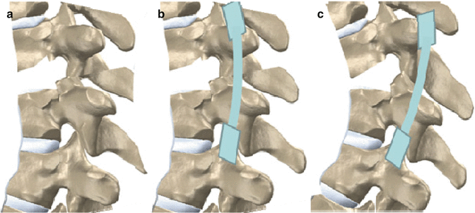
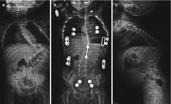
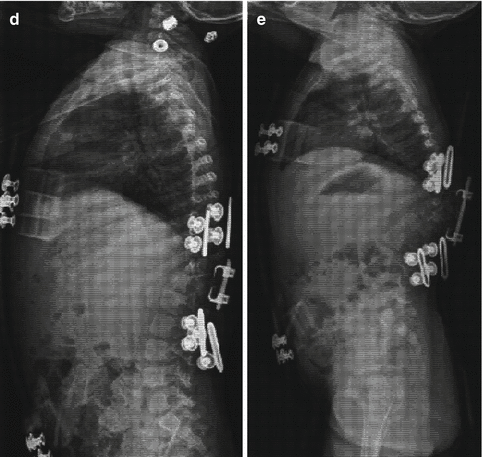
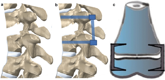
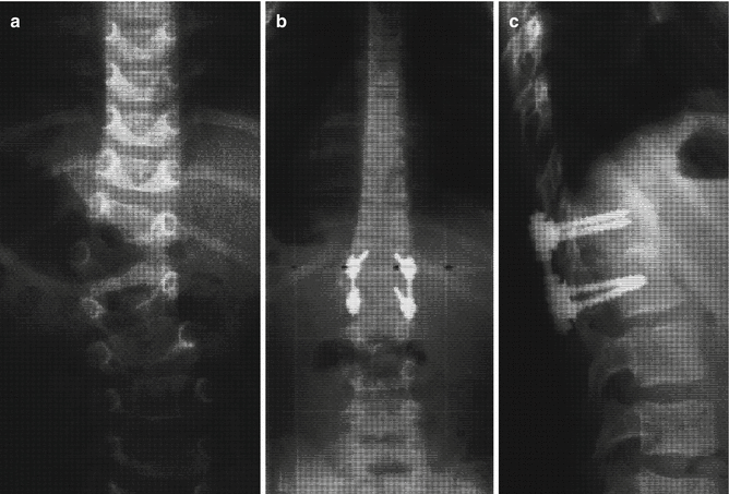

Fig. 31.5
(a) After removal of the HV. (b) Posterior compression with hooks and rods. (c) Kyphosis development (Courtesy of Gerard Bollini)


Fig. 31.6
(a) Thoracolumbar HV pre-op coronal. (b) Post-op coronal. (c) Thoracolumbar HV pre-op sagittal. (d) Post-op sagittal. (e) At follow-up sagittal with a kyphosis development

Fig. 31.7
(a) After removal of the HV. (b) Posterior compression with screws and rods construct. (c) The screws and rods construct acts like Blount staples avoiding further growth from the anterior aspect and providing stability thus avoiding kyphotic evolution (Courtesy of Gerard Bollini)

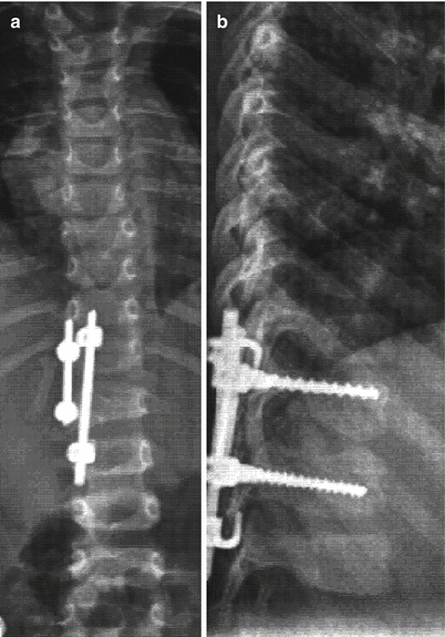
Fig. 31.8
(a) TL HV pre-op. (b) Post-op frontal view. (c) Post-op sagittal view
The Fourth Option Is to Remove Everything Between the Two Vertebral Bodies of the Vertebrae Adjacent to the HV (Fig. 31.9)
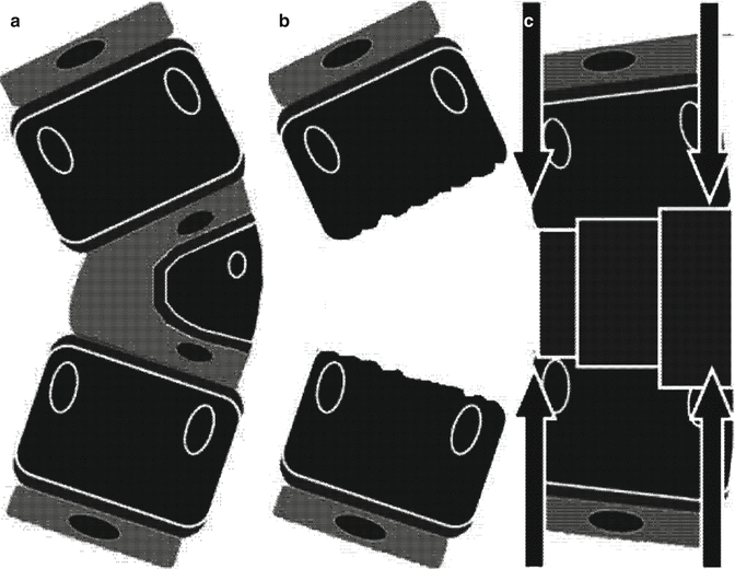
Fig 31.9
Black: osseous part of the vertebra. Dark grey: growing structures. Light grey: disc structures. (a) The HV to be removed. (b) The whole space between the two adjacent vertebral bodies is freed from all the structures. (c) Compression force is applied and the gap is filled with bone grafts (Courtesy of Gerard Bollini)
This option allows to almost complete deformity correction and total arthrodesis between the two adjacent vertebrae.
This fourth option is chosen for the older child with a markedly associated kyphotic deformity.
31.2.2 Stability Concept
Removal of a hemivertebra leaves an empty space posteriorly and anteriorly. To close the gap posteriorly and to ensure immediate posterior stability, compression instrumentation posteriorly is necessary.
Closing the posterior gap freed by the HV resection leaves an open anterior gap (otherwise you may observe a residual kyphotic deformity). This anterior gap decreases about 50 % after posterior compression. Therefore to obtain an immediate anterior stability it is necessary to use either a “screws and rods construct” or a “hooks and rods construct” associated with an anterior graft. In both cases posterior arthrodesis is mandatory.
31.2.3 Unexpected Issues
The closure of the anterior gap using a “screws and rods construct” can be tricky, in the young child, due to the small diameter of the pedicles and the fact that both the pedicle and the vertebral body are mainly cancellous bone. Loosening of the screws may occur because of the compression.
31.3 Technique
31.3.1 Preoperative Evaluation
Radiologic imaging includes standing postero-anterior (PA) and lateral view of the full spine. Magnetic resonance imaging of the spine to assess the neuroanatomy is performed to exclude intraspinal abnormalities and to study the segmentation of the HV and the growth plate. Renal ultrasound has to be done preoperatively to assess associated congenital renal system abnormalities. If the deformity looks complex on the radiographs, additional CT scan, with both 2D and 3D reconstruction, can be helpful.
31.3.2 Single Posterior Approach
The patient is positioned prone with the operative area freed for fluoroscopy. Neuromonitoring of both SSEP and MEP or NMEP is mandatory.
The pedicle of the HV is identified under fluoroscopy, the skin is marked, and a vertical midline incision is performed. The following has already been presented in Sect. 31.2.1.1.
Stay updated, free articles. Join our Telegram channel

Full access? Get Clinical Tree


