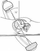
42 Roger P. Jackson and Douglas C. Burton The intrasacral (Jackson) screw and rod fixation technique involves the safe insertion of intrasacral screws and rods into the best bone possible with avoidance of injuring the surrounding neurovascular structures. The Galveston technique of intra-iliac anchorage utilizes careful osseous placement of a three-dimensionally contoured rod, taking into account spinopelvic deformity and allowing for connection to other lumbosacral anchors. Insertion of the S1 screw head into the lateral sacral mass where the head is much closer to the back of the L5-S1 disk, as well as the L5 and S1 vertebral bodies, thereby significantly reducing bending loads on the screw/rod construct at the L5-S1 level and increasing the stiffness of the fixation. The low- to no-profile rod/screw construct provides an interlocked, “sacroiliac buttress” type of fixation that allows for application of in-situ rod contouring principles to create lumbopelvic lordosis and better balance. Intra-iliac anchorage provides a strong sacropelvic foundation for long spinal constructs crossing the lumbosacral junction while maintaining a low profile in a patient population where implant prominence is often a significant concern. The bilateral placement of carefully contoured rods in the supra-acetabular intra-iliac passageway provides a strong sacropelvic foundation for the correction of rigid spinopelvic deformity and resists the flexion-extension and rotation forces that exist. This strong and stable foundation facilitates the establishment of a solid arthrodesis across the lumbosacral junction in long spinal constructs. Long spinal deformity fusions to the sacrum, spondylolisthesis, L5 burst fractures, lumbar flatback, osteo-penia or osteoporosis, and revisions including L5-S1 pseudarthrosis. Congenital abnormalities, tumors, or infections involving the proximal sacrum. Abnormalities of iliac anatomy that preclude the placement of anchorage and severe osteopenia are situations that may not allow this technique. Computed tomography (CT) scan to better delineate the sacral anatomy preoperatively. CT also is helpful in the setting of a previous or planned pelvic osteotomy, or prior bone graft harvest that has altered the normal iliac orientation for the Galveston technique. Prone positioning on a surgical table or frame that provides for improved pulmonary compliance, reduced blood loss, unrestricted fluoroscopic imaging, and that preserves or creates increased lumbopelvic lordosis, such as the Jackson surgery table. Severe hip flexion contractures and lumbar hyperlordosis are critical to recognize and account for in patient positioning. If using a standard four-poster frame, the posts must be built up to allow for the hip flexion contractures and keep undue pressure off of the knees. Hyperlordosis should be recognized preoperatively, and anterior releases performed, if needed. Positioning out of a hyperlordotic position can be aided with the use of two additional posts placed between the usual two. This eliminates, as much as possible, the sag that occurs between the posts, which accentuates lordosis. In situations with severe fixed pelvic deformity, a single femoral traction pin placed on the “high pelvis” side with 20 pounds of traction (depending on patient size) can aid in the reduction of spinopelvic deformity. Intraoperative anteroposterior (AP), lateral, and angled fluoroscopic images, by tilting of the C-arm in the sagittal plane to get a tangential view of the S1 endplate, are critical. Good AP fluoroscopic imaging of the sacrum is necessary for insertion of the intrasacral rods after the S1 screws have been deeply buried in the bone. The end of the rods should perforate the anterolateral cortex of the sacrum and not extend more than 5 to 10 mm beyond the distal aspect of the inferior SI joint. Careful preoperative planning of types and locations of anchors, particularly if S1 pedicle screws are going to be used, is paramount. If S1 screws are planned, they should be placed prior to exposure of the iliac anchor, as this will help to show how far distally the iliac anchor must be placed. If using a pedicle bolt/ connector system, short post screws are recommended, and often an open slotted connector is necessary, though not always. Transverse connection is important, and preferably performed between the S1 screws and iliac post, though anatomic considerations preclude this at times. Utilization of three- and four-rod constructs, depending on sagittal profile and coronal curve position, may facilitate Galveston rod placement. The use of a short Galveston rod on the concavity of the curve (high pelvis side) allows for ease of placement and the ability to distract against the longer, more proximally placed rod. This connection may make an S1 screw on the ipsilateral side unworkable due to bunching of connectors. A transitional lumbosacral vertebra is not a contraindication, but may make insertion of the implants more difficult, especially with partial sacralization of L5. Closely spaced iliac crests can also make the procedure more difficult. The neuromuscular patient population will frequently have severe, fixed pelvic obliquity in all three planes that makes contouring a challenge. The use of multiple rods improves, but does not eliminate, this problem. Myelomeningocele, due to its dysraphic posterior elements, widens the rod position, thereby making the connection to S1 screws and rod contouring more difficult. A good intraoperative Ferguson view of the proximal sacrum with the C-arm is needed (Fig. 42.1) A reduced-volume closed S1 screw head is deeply countersunk into the lateral sacral mass (Fig. 42.2). A special angled sacral curette is then inserted through the screw head and used to open up and sound the lateral sacral mass (Fig. 42.3). The length of intrasacral rod required can then be determined. A rod is cut to length, contoured, and directed distally into and through the closed canal of the S1 screw. Under fluoroscopic control, the rod is then worked caudally into the lateral sacral mass. The distal end of the rod is directed toward the inferior SI joint. The rod must be contoured properly and guided out laterally to get better fixation in the distal sacral mass adjacent to the SI joint. Rolling the curved rod to direct its distal end more laterally and anteriorly in the sacrum is helpful. The safest and strongest area is in the distal lateral sacrum adjacent to the SI joint. At no time is the rod intentionally driven across the sacroiliac joint. The pelvic anatomy in this area can provide considerable fixation and support for the end of the rod. This is especially so for resisting flexural bending moments. After the distal end of the rod has been driven caudally into the lateral sacral mass and out through the cortex, the proximal end is manipulated down and up into the other screw(s), then the set screw is tightened down in the S1 screw head. The second rod is implanted in the same way. Fig. 42.4 shows a typical multilevel construct with insertion of the intrasacral rods. The rod is positioned as closely to the back of the L5-S1 disk space as possible.
Intrasacral (Jackson) and Galveston Rod Contouring and Placement Techniques
Description
Key Principles
Intrasacral (Jackson) Technique
Galveston Technique
Expectations
Intrasacral (Jackson) Technique
Galveston Technique
Indications
Contraindications
Intrasacral (Jackson) Technique
Galveston Technique
Special Considerations
Special Instructions, Position, and Anesthesia
Tips, Pearls, and Lessons Learned
Intrasacral (Jackson) Technique
Galveston Technique
Difficulties Encountered
Intrasacral (Jackson) Technique
Galveston Technique
Key Procedural Steps
Intrasacral (Jackson) Technique

Stay updated, free articles. Join our Telegram channel

Full access? Get Clinical Tree


