27
Supralaminar, Infralaminar Transverse Process Hook Placement
Steven C. Ludwig
Description
The safe placement of hooks within the thoracic spine allows for surgical correction and stabilization of a variety of spinal disorders. Anchor site selections include a down-going supralaminar hook, up-going infralaminar hook, and either an up- or more commonly down-going transverse process hook.
Key Principles
Spinal instrumentation systems are characterized by a rod (longitudinal component) that is segmentally fixed to the spine via a hook or screw (anchor).
Expectations
Three-dimensional corrective forces may be delivered to the spine for deformity correction, graft compression, or spinal stabilization. Despite the merits of segmental fixation, careful attention must be directed toward the preparation of a fusion bed because all rigid implant systems will fail if a solid fusion is not achieved.
Indications
Scoliotic and paralytic deformity as well as all types of spinal instability including trauma, degenerative, neoplastic, and congenital. Laminar hooks may help shield pedicle screws and prevent late screw failure.
Contraindications
Severe osteoporosis, active posterior infection, and significant loss of anterior column stability without reconstruction. These pathologies put posteriorly placed instrumentation under excessive stress and lead to fixation failure.
Special Considerations
Careful preoperative planning is essential to determine the site of hook placement. Paired hooks in an apposing claw configuration provide a secure fixation point, especially at the ends of a construct.
Special Instructions, Position, and Anesthesia
Prone position on a variety of frame choices including Relton-Hall frame, Wilson frame, or Jackson table. Consider controlled intraoperative hypotension in the absence of significant cord compression and electrophysiology monitoring.
Tips, Pearls, and Lessons Learned
Preoperative and intraoperative communication with the surgical staff concerning the levels and types of hook facilitates the placement of the instrumentation.
Difficulties Encountered
An alternative method of segmental fixation or a change in the instrumented level should be performed if violation of the lamina or transverse process makes hook placement unsafe.
Key Procedural Steps
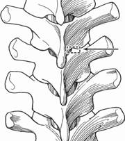
Thoracic laminar hooks can be placed against the superior or inferior edge of the lamina. Laminotomy windows are performed, using a skinny nose Leksell rongeur. Depending on the level, the interspinous/ supraspinous ligaments are taken down to expose the ligamentum flavum. At the extremes of instrumentation placement the supraspinous and interspinous ligaments should be preserved. Also, one may place hooks on each side of the spine without removing the interspinous ligaments. Following removal of the interspinous ligaments, the ligamentum flavum is removed until epidural fat is visualized. Using a 2-mm Kerrison, the laminotomy site is widened laterally. One or two millimeters of the superior facet may be removed to allow for proper hook seating (Fig. 27.1). Care is taken to bite away only the ligamentum flavum and preserve the epidural fat to avoid epidural bleeding. A trial laminar hook is grasped with a hook holder and placed into the space created. Infralaminar hooks can be directly placed under the lamina in an up-going fashion (Fig. 27.2). The blade of a supralaminar hook should be inserted by rotation about the arc of the hook to facilitate the placement and minimize spinal canal hook intrusion (Fig. 27.3). Hook purchase is checked manually with a gentle posterior-anterior maneuver.
Related posts:

Stay updated, free articles. Join our Telegram channel

Full access? Get Clinical Tree


