Dementia with Lewy bodies (DLB) and Parkinson disease dementia (PDD) share many clinical and pathologic features. They are therefore discussed together and referred to as Lewy body dementias (LBD). Whether DLB and PDD are distinct disorders, or whether they represent different presentations of the same disease, is an area of ongoing investigation. Symptoms of each disorder may arise from variations in regional and temporal onset of neural dysfunction and degeneration. By consensus, when cognitive impairments are coincident with or appear within 1 year of the motor signs, DLB is diagnosed; the term Parkinson disease dementia is used when decline occurs in the course of well-established Parkinson disease (PD). Pathologically, both conditions are characterized by the presence of Lewy bodies, intraneuronal inclusions containing α-synuclein and ubiquitin in the brain stem, limbic area, forebrain, and neocortex.
EPIDEMIOLOGY
DLB and PDD are relatively common conditions, together affecting an estimated 1.3 million individuals in the United States.
DEMENTIA WITH LEWY BODIES
DLB is considered to be a common cause of dementia, with an estimated worldwide population prevalence of 4.2% to 7.5% and incidence of 3.8% in those newly diagnosed with dementia; however, the actual prevalence and incidence rates may be much higher due to the clinical difficulty of diagnosing DLB. Men may be more susceptible to DLB and have a worse prognosis than women.
PARKINSON DISEASE DEMENTIA
The estimated prevalence of PDD among the general population is 2% to 3%. Among patients diagnosed with PD, the prevalence of dementia is more than 30%, with a lifetime incidence of up to 80% of individuals with PD who reach the age of 90 years. Although the majority of PD patients will eventually develop dementia, the time from the onset of motor symptoms to dementia varies markedly. Few prospective biomarker studies exist to assist in prediction of patients’ clinical course. Risk factors for dementia in PD include older age, age at onset of PD 60 years or older, duration of PD, and severity of parkinsonism, particularly the postural instability gait difficulty (PIGD) subtype. However, even patients with mild PD may have symptoms of cognitive impairment that does not meet criteria for dementia (mild cognitive impairment in Parkinson disease [PD-MCI]). PD-MCI may occur in up to one-quarter of patients at the time of initial PD diagnosis.
PATHOBIOLOGY
NEUROPATHOLOGY
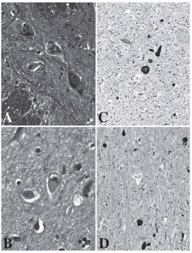
Common pathologic findings of LBD are illustrated in Figure 52.1. Lewy body (LB) pathology, particularly in the cortex, is the most important factor in the development of both DLB and PDD. There have been few comparative neuropathologic studies to determine whether the neuropathology of PDD differs from DLB or whether both conditions exist on a continuum. Neuronal loss in the substantia nigra may be more extensive in PDD than in DLB; other features including cortical LB and Alzheimer pathology have not been found to differ in PDD and DLB.
Dementia with Lewy Bodies
Pathologic features associated with DLB are summarized in Table 52.1. LBs are the only essential feature in the pathologic diagnosis of DLB; other features are apparent in most but not all cases.
LEWY BODIES
LBs are eosinophilic intracytoplasmic neuronal inclusion bodies and are found in both the subcortical regions and brain stem as well as in the cortex in DLB (see Fig. 52.1A). Brain stem LB occurs in the substantia nigra and locus coeruleus. The term cortical Lewy body refers to less well-defined spherical inclusions seen in cortical neurons (see Fig. 52.1B). In some individuals, proper identification of cortical LB may be overshadowed by the coincidence of severe Alzheimer changes; in other individuals, limitations in sampling may lead to an underidentification of cortical LB.
LEWY NEURITES
Lewy neurites (LN) are neurofilament abnormalities and a distinctive part of LB pathology in which proteins cluster into diffuse aggregates (see Fig. 52.1C and D). They occur in the hippocampus (cornu ammonis two-third region), amygdala, nucleus basalis of Meynert, dorsal vagal nucleus, and other brain stem nuclei.
ALZHEIMER PATHOLOGY
Most cases of DLB demonstrate comorbid Alzheimer disease (AD) pathology including amyloid plaques and neurofibrillary tangles. Plaque types in DLB are classified as either diffuse/immature plaques with tau-positive neurites or diffuse/immature plaques with tau-negative, ubiquitin-positive neurites.
SPONGIFORM CHANGE/MICROVACUOLATION
This is a feature of some DLB cases and occurs mainly in temporal cortex. It may relate to the severity of the disease. Although similar to spongiform change in Creutzfeldt-Jakob and prion-related diseases, there is no evidence that DLB is a transmissible disorder or linked to abnormal prion protein.
Parkinson Disease Dementia
Although PD often coexists with other common causes of dementia, such as AD, neuropathologic studies have found that the degree of LB pathology (including LB and LN) may have a closer correlation with cognitive decline and dementia in PD than does the degree of AD pathology.
FIGURE 52.1 Pathology of Lewy body dementias. A: Pars compacta of the substantia nigra: two pigmented neurons each one including two Lewy bodies. Subcortical Lewy bodies have a basophilic central core or are eosinophilic depending on the plane of section and are surrounded by a pale, narrow halo. B: Occipitotemporalis gyrus, fifth layer including a cortical Lewy body-containing neuron. Cortical Lewy bodies are ill defined and diffusely eosinophilic. The nuclear chromatin of cortical Lewy body-containing neurons is often vesiculated. C: Nucleus coeruleus. D: Substantia innominata or nucleus of Meynert. Both C and D exhibit α-synuclein-labeled Lewy bodies and Lewy neurites. (A and B, Luxol fast blue counterstained with hematoxylin and eosin; C and D, α-synuclein. Original magnification: A, 400×; B, 630×; C and D, 200×. Courtesy of JP Vonsattel, MD.)
A possible synergistic role between AD and PD pathology needs further research. A few studies have found that reduced amyloid β1-42 in the cerebrospinal fluid is associated with cognitive decline in patients with PD, as it is in patients with AD alone. Other data suggest that that α-synuclein deposition promotes the intracellular aggregation of tau and β-amyloid in cell models and that there is a more rapid disease progression in PDD cases with coexistent AD. It is likely that given the substantial prevalence of both AD and PD, at least some patients with PD may have cognitive decline that is attributed to or exacerbated by AD.
NEUROTRANSMITTER SYSTEMS
The exact mechanisms leading to dementia in DLB and PDD are unknown. However, there is an association between structural neuropathologic findings in LBD; dopamine depletion; and noradrenergic, serotonergic, and cholinergic dysfunction. LBs have a predilection for the substantia nigra, and both PDD and DLB are characterized by dopaminergic changes in the striatum and frontal cortex, leading to parkinsonism. In addition to motor control, dopamine is involved in a range of nonmotor behaviors such as cognition, motivation, sleep, and mood. The cholinergic nucleus basalis is another LB predilection site, leading to widespread and severe cortical cholinergic deficits. LBD pathology may also affect brain stem nuclei such as the locus coeruleus, which receives noradrenergic activation, and the serotonergic raphe nuclei. All of these areas are known to be involved in modulation of cognitive and psychiatric symptoms.
GENETIC RISK FACTORS
Although large genome-wide association studies (GWAS) have identified a variety of genes contributing to sporadic PD, similar large studies have not been published yet for PDD, and only one GWAS study has been done in DLB. However, the importance of genetic factors for these diseases has begun to be established.
TABLE 52.1 Pathologic Features Associated with Dementia with Lewy Bodies
Essential for diagnosis of DLB:
Lewy bodies
Associated but not essential for diagnosis of DLB:
Lewy-related neurites
Plaques (all morphologic types)
Neurofibrillary tangles
Regional neuronal loss, especially in the brain stem (including the substantia nigra and the locus coeruleus) and the nucleus basalis of Meynert
Microvacuolation (spongiform changes) and synapse loss
Reduced concentrations of choline acetyltransferase (ChAT) in neocortex
DLB, dementia with Lewy bodies.
Glucocerebrosidase
Several individual studies and an international multicenter collaborative study have demonstrated that patients with PD who carried mutations for the lysosomal enzyme glucocerebrosidase (GBA1 ) have a higher incidence of PD-MCI and PDD. Other multicenter autopsy studies have demonstrated that subjects with DLB were more than eight times more likely to carry a GBA1 mutation than were controls. Further analyses indicate that GBA1 mutations are also significantly associated with earlier age at onset and death in DLB. Although mechanisms underlying GBA1-associated parkinsonism and dementia are still not completely understood, accumulating evidence suggests that impairment of the aging lysosome, enhanced by deficient or mutant glucocerebrosidase, can affect α-synuclein degradation.
Leucine-Rich Repeat Kinase 2
To date, cross-sectional studies have not identified specific cognitive dysfunction associated with mutations in leucine-rich repeat kinase 2 (LRRK2), a common cause of genetic PD. Of note, LRRK2 G2019S carriers are more likely to have the PIGD phenotype, which is more likely to be associated with cognitive impairment. The PIGD phenotype, however, has not been associated with cognitive impairment in cross-sectional studies of LRRK2 G2019S carriers.
Apolipoprotein E
Although cross-sectional studies have yielded disparate results, two prospective cohort studies found no association between APOE ε4 genotype and the development of PDD or rate of change on the MMSE in patients with PD. One study examined rate of change on the Mattis Dementia Rating Scale (MDRS) over time and showed that the ε4 allele was associated with a more rapid decline in MDRS scores (3 points per year; hazard ratio 2.8). In contrast, a recent large multinational case-control genetic association study using pathologically diagnosed samples demonstrated that the APOE ε4 allele was a strong genetic risk factor for DLB.
CLINICAL MANIFESTATIONS
DEMENTIA WITH LEWY BODIES
The core diagnostic criteria for probable and possible DLB as proposed by the third report of the DLB consortium, and as commonly used in clinical practice, are included in Table 52.2 [Level 1].1 These core criteria have a sensitivity of 83% and a specificity of 95% for the presence of neocortical LBs at autopsy. However, these core criteria are more predictive of the relatively rare “pure” form of DLB rather than the more common findings of mixed DLB and AD pathology.
Supportive clinical features include repeated falls, syncope, transient loss of consciousness, severe autonomic dysfunction, depression, systematized delusions, or hallucinations in other sensory and perceptual modalities. The presence of low uptake in a myocardial scintigraphy study has been included as a supportive feature in recently revised criteria for diagnosis of DLB. Although these features may support the clinical diagnosis, they lack diagnostic specificity of the core features and can be seen in other neurodegenerative disorders.
PARKINSON DISEASE DEMENTIA
The diagnosis of PDD requires a clinical diagnosis of PD accompanied by cognitive impairment of sufficient magnitude to interfere with social or occupational function and the criteria for diagnosis as outlined by the International Parkinson and Movement Disorder Society are presented in Table 52.3. Criteria for diagnosis of PD-MCI have also been established (Table 52.4). It is estimated that PD-MCI occurs in up to 25% of nondemented individuals with PD and predicts a more rapid cognitive decline and shorter time to dementia.
TABLE 52.2 Diagnostic Criteria for Dementia with Lewy Bodies
Core features of DLB
• Fluctuating attention and concentration
• Recurrent well-formed visual hallucinations
• Spontaneous parkinsonism
Suggestive features of DLB
• Rapid eye movement (REM) behavior disorder
• Severe neuroleptic sensitivity
• Low dopamine transporter uptake in the basal ganglia demonstrated by ioflupane I-123 dopamine transporter SPECT imaging (DaTscan)
Probable DLB: dementia plus
Two core features OR
One core feature AND one suggestive feature
Possible DLB: dementia plus
One core feature OR
One suggestive feature
DLB, dementia with Lewy bodies; SPECT, single-photon emission computed tomography.
Data from McKeith IG, Dickson DW, Lowe J, et al. Diagnosis and management of dementia with Lewy bodies: third report of the DLB Consortium. Neurology. 2005;65:1863-1872.
Only gold members can continue reading. Log In or Register to continue