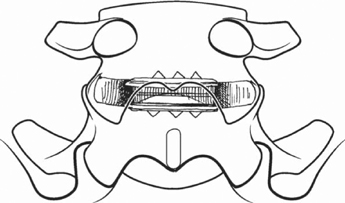
78 Scott L. Blumenthal Spine fusion for degenerative disk disease has been regarded as the gold standard, but this has not produced excellent results, with pseudarthrosis being an all-too-common occurrence, as well as patients who achieve a solid fusion but who do not have a favorable clinical outcome. Spine fusion surgery has also given rise to the phenomenon of adjacent segment disease related to the relatively rigid fusion segment being adjacent to a previously normal segment. These concerns have highlighted the need for a surgical treatment that spares spinal motion, allowing motion to be closer to that of pre-illness levels and to allow a more rapid return to work and other activities without having to rely on a solid fusion first taking place. It is essential first to identify the cause of the low back pain. Disk replacement should not be considered in patients with fracture, infection, spondylolisthesis, spinal stenosis, sacroiliac problems, or facet-related pain. These diagnoses should be ruled out as the primary pain source. History and physical examination followed by anteroposterior (AP) and lateral flexion and extension radiographs should be performed. Magnetic resonance imaging (MRI) is a helpful study to evaluate the intervertebral disks, spinal canal, and neural foramen. One must be cautious in assuming that every degenerated “black disk” found on MRI is a source of pain. We have found it helpful to perform a diskography to evaluate intradiskal pressure and volume, disk morphology, and most importantly, reproduction of the patient’s usual pain during disk injection. Pain intensity during injection can be assessed on a 0 to 10 visual analogue scale (VAS) at each level. The patients should be awake and blinded to the level being tested. Facet pain may be ruled out by history and physical examination. If in doubt, a facet injection may be performed. Bone densitometry should be obtained on all patients being worked up for a spinal arthroplasty procedure, especially if there is any concern about patients’ bone quality or if their history indicates a high risk of osteoporosis. Pain arising from degenerative disk disease at one or more lumbar levels not responding to nonoperative measures is the primary indication for spinal arthroplasty. The goals of spinal arthroplasty are to maintain motion, relieve pain, preserve disk space height, maintain neural foraminal height, and preserve the facet joints. It is important to have longevity of any implant through millions of cycles and to aim for avoidance of revision surgery. This requires excellent wear characteristics of any spacer, excellent shock absorbing capacity, and a solid interface between the device and the host bone without sacrificing revision options. The expectation of the surgical procedure is that patients experience significant relief of their back pain complaints and are able to resume activities of daily living as well as some recreational activities or a preinjury occupation. It is hoped that the long-term potential of symptomatic adjacent level disease is minimized with the placement of a motion-sparing device rather than a spinal fusion. Infection and instability must be ruled out as well as low bone density, typically defined as a density value one standard deviation below the normal population mean. Typically, patients may be no older than 60 years of age. Facet arthrosis at the level being considered may be present but should not be severe. The presence of radicular pain in addition to axial pain is acceptable if there is no severe canal stenosis and it is thought that the radicular pain is mostly from foraminal stenosis secondary to loss of disk height at that level. This may be corrected with the distraction that is performed during insertion of the artificial disk. The presence of infection or disruption of posterior structures or any instability from a pars defect or pedicle fracture should be regarded as absolute contraindications. The presence of a rigid deformity is a contraindication. Bending films may be obtained to differentiate sciatic scoliosis from other rigid forms such as degenerative or idiopathic. Most artificial disk devices consist of a metal end plate that anchors into the vertebral endplate (Fig. 78.1). These metal end plates articulate with a spacer (Fig. 78.2), which allows motion at the segment (Fig. 78.3). The metal end plates are available in different cross-sectional sizes and lordotic angles, and come with or without a porous coating and have either teeth or keels to gain stability into the bony vertebral end plate. The spacers come in various thicknesses and consist of either metal or polyethylene (Fig. 78.4). The total disk replacement (TDR) devices have varying levels of constraint.
Lumbar Spinal Arthroplasty
Description
Key Principles
Expectations
Indications
Contraindications
Special Considerations

Stay updated, free articles. Join our Telegram channel

Full access? Get Clinical Tree


