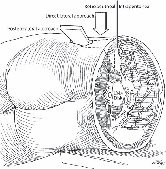
67 Luiz Pimenta The extreme-lateral lumbar interbody fusion (XLIF) procedure is a minimally disruptive direct-lateral, retroperitoneal, trans-psoas approach to the lumbar spine. It incorporates blunt finger dissection of the retroperitoneal space, stimulated electromyogram (EMG) guidance of blunt dilators through the psoas muscle, and advancement of a split-blade retractor system for illuminated direct visualization of the lateral portion of the spine. The approach is described here for interbody fusion, but has also been used as a revision strategy for failed anteriorly placed total disk replacement and as a primary procedure for laterally placed total disk replacement. The direction of the approach avoids the muscle, bone, and ligament disruption of a posterior approach and the vascular and visceral mobilization and potential concomitant risks of an anterior approach. The minimal dissection and tissue damage result in quick operative times, minimal blood loss, minimal morbidity, and quick patient recovery. The lateral exposure allows for more complete disk space preparation and optimal placement of a large interbody device, which, when coupled with preservation of both anterior and posterior longitudinal ligaments, provides for an exceptionally stable construct. Working in a lateral position through the retroperitoneal anatomy may feel foreign to those accustomed to the more traditional anterior or posterior approaches. However, because the exposure procedure itself is relatively simple, and the intervertebral disk work is conventional, the learning curve is not steep. XLIF can be performed without the assistance of an approach surgeon in about an hour, with barely detectable blood loss. Disk height, foraminal volume, and spinal alignment can be superbly achieved using a large implant, positioned optimally in the anterior half of the disk space and sitting along the biomechanically sound ring apophysis. Patients tend to have few postoperative complaints, aside from a transient hip flexor pain or weakness (depending on how delicately the psoas muscle is dissected), and can generally ambulate within hours of surgery and be discharged from the hospital the next day. The indications for XLIF fusion surgery are those for any L1-L5 interbody fusion, and include, but are not limited to, degenerative disk disease with instability, recurrent disk herniation, low-grade degenerative spondylolisthesis (less than grade 3), degenerative scoliosis, adjacent level syndrome, postlaminectomy instability, and posterior pseudarthrosis. The XLIF surgery, as a stand-alone procedure, relies on indirect decompression of the neural elements from restoration of disk height and spinal alignment, but the need for direct decompression does not necessarily contraindicate XLIF. Performing a conjunctive posterior microdecompression adds minimally to the surgical time and retains the minimally disruptive benefits of the procedure. Contraindications include those for interbody fusion surgery in general, such as infection, unless an autograft is placed, or there are significant comorbidities. Interestingly, although osteoporosis may contraindicate other fusion procedures, low bone quality is better tolerated by the XLIF procedure due to the size and position of the interbody implant. The direct lateral approach is specifically contraindicated at L5-S1 due to the location of the iliac crest, and in patients with bilateral retroperitoneal scarring, such as from prior surgery. Although degenerative scoliosis is a growing application for the XLIF approach because of the potential for alignment correction and low morbidity for a typically elderly population, lumbar deformities with greater than 30 degrees of rotation can confuse the approach and present risk. Similarly, high-grade degenerative spondylolisthesis (higher than grade 2) may require more demanding techniques for slip reduction. Large patients tend to be more difficult than thin patients from most approaches. However, in XLIF the patient is in a lateral decubitus position, which allows the abdomen to fall anteriorly away from the spine, shortening the distance from skin to spine (which is more dependent on the width of the hips than the girth of the waist), and opens the retroperitoneal space to provide a large safe approach trajectory. In all patients, it is important to consider the nerves of the lumbar plexus within the psoas muscle, which typically lie in the posterior third of the muscle. Given variation in anatomy and surgical approach, care is warranted and EMG-assisted nerve avoidance is recommended. Patient positioning is key to an unencumbered successful XLIF procedure. The patient should be positioned on a radiolucent breakable surgical table in a true lateral decubitus position. A true lateral position should be confirmed with fluoroscopy prior to taping and draping and reconfirmed periodically. It is easiest if the patient or surgical table is positioned to provide a true lateral, such that the fluoroscope C-arm will be used at a 0- or 90-degree angle for lateral or cross-table anteroposterior (AP) images only, rather than adjusting the angle of the C-arm to fit the angle of the patient. This helps to ensure that access exposure and instrument manipulation occur in the true lateral direction, maximizing implant placement and minimizing risk to anterior or posterior structures. The patient should be securely taped to the table in this true lateral position, with the iliac crest at the break in the table, and the tape securely leveraging motion of the iliac crest inferiorly away from the waist. Breaking the table then at the crest provides for added working space between the rib cage and the ileum and facilitates exposure, especially at L4-L5. The patient’s hips should be flexed to relieve tension on the psoas muscle. For EMG nerve monitoring during the trans-psoas approach and during interbody distraction and implant insertion, anesthesia should not include muscle relaxants or paralytic agents subsequent to initial shortacting agents given for intubation, or they should be reversed prior to the start of the approach. Four out of four twitches from a train-of-four test is required. A good fluoroscopic image is extremely helpful. Adjust the cephalocaudal angle of the C-arm to provide a clear lateral view of the end plates. There is a tendency to cheat the initial exposure anteriorly to avoid the nerves posteriorly. However, because the XLIF retractor is designed to prevent pressure on the posterior elements, the aperture is preferentially expanded anteriorly. Therefore, the ideal initial target spot is the direct center of the lateral aspect of the disk, which will result in retractor exposure of the anterior half of the disk space. Multilevel procedures can be performed using the same skin incision but separate fascial incisions and psoas muscle dilations. In degenerative scoliosis cases, coronal alignment can be achieved from either side, but access is easier from the convex side because of osteophyte formation on the concave side. The contralateral annulus must be disrupted to achieve parallel distraction, optimal biomechanical position of the implant, and optimal coronal alignment. Symmetrical cephalocaudal expansion of the retractor can be deflected by the iliac crest at L4-L5 or by the ribs at L1-L2 or L2-L3. Access difficulty at L4-L5 can be overcome with proper positioning and aggressive bending of the patient. At L2-L3, the 12th rib may need to be retracted or partially resected. At L1-L2, an intercostal approach may be necessary between the 11th and 12th ribs. If nerve monitoring warns of a too posterior position within the psoas, slight translation of the dilators away from the indicated direction of the nerve(s) should still provide adequate exposure. A stimulating ball-tipped probe can be used to identify tissue structures within the exposure if necessary. Proper targeting of the disk space should prevent a too anterior exposure, but care should be taken to limit retractor excursion or disk work to posterior to the anterior longitudinal ligament. Lateral fluoroscopic images should be obtained to ensure AP position.
Minimally Disruptive Lateral Approach to the Lumbar Spine: Extreme-Lateral Lumbar Interbody Fusion
Description
Key Principles
Expectations
Indications
Contraindications
Special Considerations
Special Instructions, Position, and Anesthesia
Tips, Pearls, and Lessons Learned
Difficulties Encountered
Key Procedural Steps

Stay updated, free articles. Join our Telegram channel

Full access? Get Clinical Tree


