4 Nasal Endoscopy in the Preoperative Assessment
Juan Eugenio Salas Galicia, Raúl Omar Cadena Torrero, and María Chávez Méndez
 Introduction
“A picture is worth a thousand words.” The images provided by endoscopic examination are a powerful diagnostic tool in the area of otorhinolaryngology–head and neck surgery and neurosurgery, particularly for the evaluation of the nose, paranasal sinuses, and skull base region. Nasal endoscopy was first performed by Hirschmann in 1901 using a modified cystoscope, and soon after it was applied in minor surgical procedures by Reichert, Valentine, Sargnon, and Zaufal, from 1902 to 1908. The use of endoscopes for diagnostic and surgical treatment purposes presented a great development in the late 1960s with the introduction of the Hopkins cylindrical lenses, thereby improving the diagnostic and surgical capabilities of this instrument both for providing a direct vision and for photo documentation. Messerklinger introduced systematic exploration of the lateral nasal wall, confirming with his vast clinical experience that most of the diseases of the paranasal sinuses are rhinogenic. He also found that these diseases were generally related to two fundamental areas that he called the ostiomeatal complex and the sphenoethmoidal recess. His studies provided more precise knowledge of the anatomical structures and the sinonasal physiology.1–3
 Indications
Diagnostic nasal endoscopy is indicated in the following cases:
1. Chronic obstructive rhinopathy
2. Recurrent or chronic rhinosinusitis
3. Facial neuralgia or headache, mostly associated with previous sinonasal surgery
4. Persistent rhinorrhea
5. Epistaxis
6. Epiphora
7. Chronic pharyngitis or laryngitis
8. Rhinopharynx diseases
9. Chronic or recurrent otitis media
10. Anosmia or hyposmia
11. CSF leakage (topodiagnostic)
12. Sinonasal tumors
13. Skull base tumors
14. Sinonasal and skull base tumor biopsies (when indicated)
15. Postoperative follow-up of endoscopic sinus surgery and transnasal craniectomy2,4–6
 Contraindications
Rigid nasal endoscopy in the doctor’s office under local anesthesia is usually contraindicated in uncooperative children or in apprehensive adults or psychiatric patients. For these patients, flexible endoscopy under topical anesthesia is well tolerated. General anesthesia is rarely required.
 Risk Factors
In general, nasal endoscopy should be avoided in the elderly and in patients with a history of asthma and hypersensitivity, heart disease, and facial pain, and in hypervascularized tumors when their size makes it difficult to introduce the endoscope into the nasal cavity.
 Complications
Complications of nasal endoscopy in the doctor’s office are very uncommon and may occur due to vasoconstrictors and local anesthetics, which can occasionally cause tachycardia, hypersensitivity, and high blood pressure. Other complications are related to the procedure itself, and include injury to the mucosa, epistaxis, vagal reaction, nasofacial neuralgias, and asthmatic events. After taking into account the contraindications and risk factors listed above and these possible complications, nasal endoscopy, as a preoperative evaluation, is a safe and effective procedure of high diagnostic value.2,3,5,6
 Equipment
Telescopes: 2.7 mm, 30, 45, and 70 degrees; 4 mm, 30 and 45 degrees; flexible nasofibrolaryngoscope, 3.5 mm/ 2.5 mm in diameter
Light source: Xenon 175 or 300 W; cold light source LED Nova 100 W (new option)
Cable of fiber optics or optic fluid (recommended for photo documentation)
Video: Analog or digital, three chips or high-definition camera (HD Image 1, Karl Storz Endoscopy, Flanders, NJ); the authors consider the high definition camera the most appropriate in terms of image
Monitor: A 19″ LCD flat monitor SXGA (1280 × 1024) resolution or a 23″ wide-view HD monitor (1920 × 1200) resolution11
Documentation: HDD and DVD video recorder or Blue-Ray HDD recorder (Sony or Panasonic); UP-D 55 digital Sony video printer
 Computer Equipment
Hardware: PC: Intel dual or quad core processor; Chipset Intel motherboard; DDR2 or DDR3 memory, 4.0 Gb recommended; S-ATA-II hard disk drive, 300 MBps, 7200 Rpm, 320.0 Gb recommended; HD or full-HD capable PCI express 16× video card, 512 Mb DDR2 or DDR3 minimum; LCD monitor 20″ wide screen with digital inputs (DVI or HDMI); DVD or Blu-Ray writer drive; multiple memory flash card reader; PS2 or USB keyboard; PS2 or USB laser mouse; uninterruptible power system, with at least twice the total power consumption. A mirror disk array (Raid-1) is recommended for data protection in case of hardware failure.
Macintosh HD: Intel Xeon “Nehalem” dual or quad core processor, 2.66 GHz, 3 to 8 GB memory, 640 GB hard disk; Express 18— super drive disc; Chipset motherboard, NVIDIA GeForce GT 120 with 512 MB; LED Cinema Apple Monitor, 24″ display, 1920 × 1200p image
PC software: Windows XP Professional is the most stable operating system (with the Pinnacle studio 12 capture and editing software).
Mac software: XO 10.5.6 operating system with the Final Cut Pro Edition software
Data archiving system: The Karl Storz AIDA® DVD-M is the ideal system (state of the art) for data, image, and video archiving, report writing and printing, and automated storing with image or video inspection, DVD, CD-ROM, or USB stick recording. Another database solution for file control is Capturis, a powerful database developed on FileMaker Pro 10; it works on a PC or Mac platform (Fig. 4.1). Other options on the market are Pentax-Kay, Olympus, Ecleris, and others. Clinicians must choose a system that is appropriate for the particular needs of their daily practice.
 Method
Basic Diagnostic Nasal Endoscopy
In addition to nasal endoscopy, several functional tests may also be used to evaluate the nasal cavity, including active anterior rhinomanometry and acoustic rhinometry (Fig. 4.2), olfactometry, and mucociliary clearance. It is important to avoid using irritating substances that can disturb the nasal mucosa. The diagnostic nasal endoscopy is performed with the patient seated or in a recumbent position, after a traditional anterior rhinoscopy (Fig. 4.3). The evaluation takes place without any anesthesia or vasoconstriction medication. The nasal cavity is assessed with a 2.7-mm, 30-degree endoscope, which offers a wider anterior view. The tip of the endoscope is inserted slowly and carefully into the vestibule and nasal valve region to avoid any discomfort to the patient, without touching the nasal septum or the lateral nasal wall. It is very important to visualize both nasal cavities as completely as possible. The characteristics of the mucosa, mucus, and nasal secretions are assessed, as well as the anatomical variations. This procedure is more difficult to perform with a 4-mm telescope due to its larger diameter. In this phase, cultures guided by endoscopy may be taken by using a suctioncollector device (Juhn Tym-Tap®, Xomed Inc., Jacksonville, FL) or even with an ear aspirator. A cheaper alternative would be a urologic swab for culture.
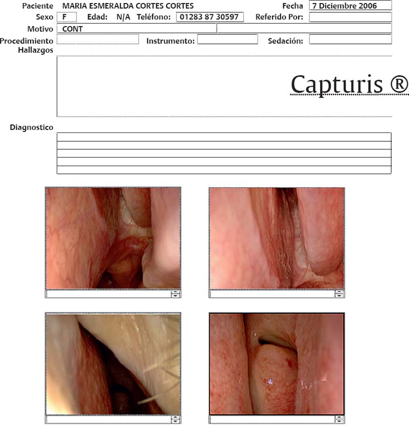
Fig. 4.1 Endoscopic Capturis report sheet.
Related posts:
 Transmaxillary Endoscopic Approach to Contralateral Parasellar Lesions


