Fig. 28.1
Idiopathic EOS. Scoliosis was noted at the age of 6 years and progressed to 30° by the age of 7 years (a). Full-time brace treatment began at the age of 7 years (b) and continued through the age of 13 years, then part-time at the request of the patient. At follow-up at the age of 18 years after 1 year out of brace, there is a stable 25° curve (c)
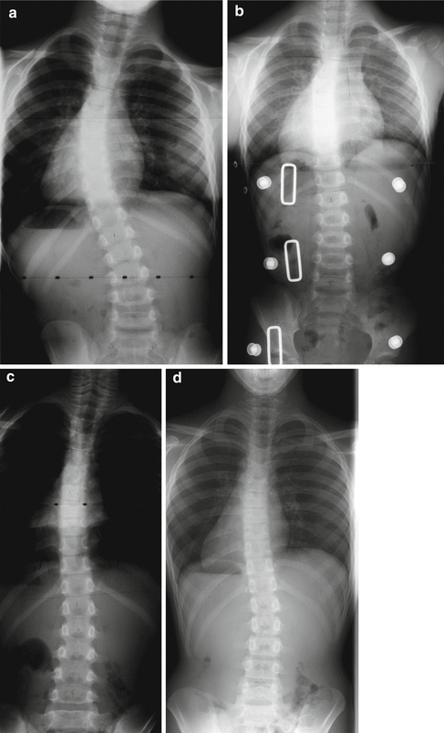
Fig. 28.2
Idiopathic early onset scoliosis. Scoliosis noted at the age of 3 ½ years (a) and full-time bracing initiated with good in-brace correction (b). At the age of 6 years, correction was maintained (c). Part-time bracing and correction were maintained through adolescence (d)
Success or failure in bracing depends partly upon the goals chosen for treatment. Establishing realistic, specific, and transparent goals early in orthotic treatment of early onset deformity facilitates rational expectations by the practitioner and family. Is the goal complete correction, prevention of worsened deformity, or slowing of progressive deformity, acknowledging that surgery will eventually be needed? Complete, lasting correction with repetitive casting is a reasonable goal in early, selected idiopathic scoliosis as demonstrated by Mehta [10], and is anecdotally occasionally also achieved in moderate EOS in the older age range treated by bracing alone (see Fig. 28.2). Complete correction is rarely achieved in progressive idiopathic scoliosis in the younger age range by orthotic treatment alone and repetitive casting may be a better choice. Complete correction as a goal can help motivate families and patients assuming there is some chance of achieving that goal. Bracing is usually used in conjunction with casting even when complete correction is obtained, and the brace is used to maintain this correction after the cast is discontinued. In more severe EOS where complete correction is unlikely, nonoperative treatment is sometimes viewed as a temporizing measure, stabilizing deformity for years, allowing for more growth before initiating surgical treatment, and allowing the child to remain free of the need for repetitive surgical intervention with growing rods [11] (Fig. 28.3). Nonoperative treatment to allow for more growth with eventual surgery anticipated can be successful in achieving the dual goals of a longer spine and fewer operations, but also may lead to inappropriate delay and worsened, irrevocable, thoracic deformity (Figs. 28.4 and 28.5). The availability of modern growth friendly surgical treatments for spinal deformity such as expandable spinal rods or VEPTR should lower the threshold for discontinuance of bracing and initiation of surgery to a point before spine or chest deformity become too severe. Unfortunately quantifying the appropriate time for discontinuation of bracing is not defined, but is often determined by monitoring change of chest shape and spine deformity on serial radiographs and physical exam.
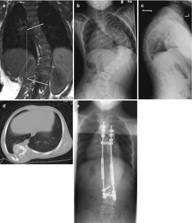
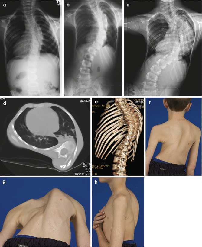
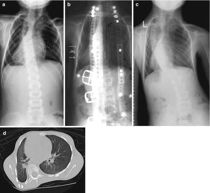

Fig. 28.3
Idiopathic early onset scoliosis. Treatment began at 18 months with full-time brace treatment (a). Referred for growing rods at the age of 7 years when rib prominence and thoracic deformity had worsened (b–d). Spine and thoracic deformity have been fairly well-controlled by dual growing rods from the age of 7 to 11 years (e). Growing rods had been suggested at the age of 2 years, but declined by the family. Earlier surgical intervention would have resulted in an easier initial surgical procedure, but the child was spared 5 years of surgical interventions and 10 surgical lengthening procedures by brace treatment from the age of 18 months until 7 years

Fig. 28.4
Idiopathic early onset scoliosis treated with repetitive casting and bracing beginning at the age of 2 years. Initial thoracic deformity at the age of 3 years (a) was modest, worse at the age of 5 years (b), and severe at the age of 8 years (c), with pulmonary function tests approximately 50 % of predicted and early restrictive lung disease. The convex thorax is collapsed, with the ribs assuming a vertical orientation or “collapsing parasol deformity” as described by Campbell. Thoracic deformity is demonstrated on CT (d, e). An area of the posterior convex thorax normally occupied by lung is occluded. Thoracic deformity is clinically apparent (f–h). Spinal deformity can be controlled surgically at this stage, but the chest deformity will not be completely improved by surgical means. Earlier intervention with a growth-oriented technique such as dual growing rods or VEPTR might have controlled both spine and chest deformity and led to a better result

Fig. 28.5
Congenital early onset scoliosis in the upper thoracic spine was treated with circumferential in situ fusion at the age of 2 years (a). The normally segmented curve below was then treated with a full-time Milwaukee brace with a pad pressure applied (arrows) directly laterally over the convex chest wall (b). At the age of 9 years, the child was referred for surgical treatment because of curve progression (c) while still using the brace. Chest deformity with a severe collapse of the convex chest wall (d), however, has been evolving for years and is now irrevocable. Earlier growing rods would have been a better choice than persistent brace management
Effective brace treatment of early onset scoliosis demands appropriate indications, practical expectations, an effective brace, and committed care-givers. Commitment to bracing at the level of the physician (and the rest of the medical team), orthotist, and family is critical for success. Absence of dedication by any of the team will subvert the efforts of the others. Creating an effective scoliosis orthosis requires a skilled orthotist with experience in treating EOS patients. Although well-documented bracing systems of many types are available, it is difficult to be successful in early onset scoliosis without experience at some level. Not all techniques applicable to adolescents are transferable to early onset scoliosis age group. Fortunately, the appropriateness of the specific orthosis and its potential effectiveness is easily assessed by radiographs taken in the brace. Radiographic confirmation of the effectiveness of bracing should complement clinical examination of the chest wall deformity. Patient compliance with requested brace usage is a major barrier to success in adolescents but much less problematic in younger children with early onset deformity if families are committed to brace wear. Often, the most likely member of the team to lack commitment is the physician, who may imply “try this brace for a while, it probably won’t work; come back and see me when you need an operation.”
28.2 Evidence for Efficacy of Bracing in Idiopathic Early Onset Scoliosis
Mehta’s [10] experience with casting for idiopathic EOS is now well documented and shows remarkable, lasting correction of many patients in whom treatment was begun early and even some long-term improvement in many in whom referral was late. This experience clearly shows that the deformed growing spine can be guided through growth not just with stabilization of deformity but with actual long-term improvement in deformity, and that complete correction may be possible if the early infantile growth rate is harnessed to curve correction. The obvious advantage of casting includes full-time use without the need for adherence to bracing regimens. Mehta’s [10] series included patients up to 48 months in age and her experience is relevant to bracing of early onset curves as it shows convincingly that with growth and appropriate application of external pressure, the deformed growing spine can be changed for the better. Mehta [12] has also advocated the use of serial plaster casts in older patients with idiopathic EOS, but this is less well documented. Experience with brace treatment alone for younger patients with idiopathic scoliosis is sparsely reported [12–17]. McMaster and Macnicole [18] documented Milwaukee brace treatment in 27 children with idiopathic EOS in young children, of whom only 5 did not require surgery during adolescence. However 70 % of the children in the study wore the brace a minimum of 5 years suggesting that bracing delayed the need for surgical intervention.
Experience reported with brace treatment of older patients with idiopathic EOS is encouraging. With notable exceptions [12, 19–24], this experience is blended into reports on success or failure with adolescent idiopathic scoliosis. Robinson and McMaster [24], in analyzing curve patterns in idiopathic EOS, reported 88 of 109 patients who were treated with a brace. Arthrodesis was needed in 67 of 84 thoracic curves but in only 3 of 20 thoracolumbar or lumbar curves. However, the mean Cobb angles were higher in the thoracic curve patterns at the initiation of treatment [25, 26] compared to the thoracolumbar and lumbar curve patterns [22, 27]. Curve correction in brace was best below the age of 6 years and early in bracing. Noonan et al. [28] discouraging report of bracing in idiopathic scoliosis included patients as young as the age of 8 years, but EOS patients are not distinguished from the rest, although the authors noted a higher failure rate in patients under the age of 12 years. The experience with the Boston Brace system in 295 patients [29] at our institution included 34 patients in the age of 10 years or less. When compared with adolescents in the study, patients less than 10 years old at initiation of bracing had a higher rate of surgery, but also a higher mean correction at the end of bracing in those who did not need surgery. There were very few patients whose curves remained the same by the end of growth, probably reflective of the large amount of growth and opportunity for change in either direction during treatment. We felt that bracing as a whole for this group was successful. Mean curve correction at the end of bracing was 25 % for patients who were successfully treated with bracing. Of all patients between the age of 4 and 10 years at initiation of brace treatment, only 5 of 34 went on to surgery. Of those less than 10 years of age starting bracing with curves between 30 and 49°, only 2 of 11 went on to surgery. Tolo and Gillespie [30] reported on 44 patients braced for EOS, among whom, 16 went on to surgery and felt that part-time brace use might be effective. Jarvis et al. [20] reported on 23 patients and also felt part-time bracing was effective. Kahanovitz et al. [27] reported on treatment of 15 EOS patients with part-time bracing and noted success for patients who had curvatures less than 35° at the onset of part-time bracing and whose rib vertebrate angle difference remained less than 20°.
All these studies suffer from being retrospective selective reviews without either cohort controls or prospective controls. With the exception of Noonan, however, all observed encouraging outcomes for patients treated with braces. The Scoliosis Research Society prospective study of bracing in idiopathic scoliosis by Nachemson and Peterson [31] demonstrated efficacy in bracing for adolescent idiopathic scoliosis, but does not include patients below the age of 10 years. In a recent study by Weinstein et al., the rate of treatment success with bracing was 72 % after bracing, as compared with 48 % after observation in a prospective study of patients aged 10–15. This study also demonstrated a dose response with regard to hours of wear, but again whether these findings translate to the early onset population is not known [8]. In addition, the rate of successful treatment by observation or bracing was worse in young patients who were Risser 0. In Risser 0 patients, the calculated probability of failure ranged from 32 to 91 % depending on the initial Cobb angle compared to 7–53 % depending on the initial Cobb in Risser 1+ patients. Bracing reduced these probabilities to 13 % and 76 % and 2 % to 28 % with bracing, respectively [32].
Long-term studies of outcome after bracing for adolescent idiopathic scoliosis [33, 34] indicate a favorable long-term result with regard to pain and function. All show a cohort of patients functioning well with no report of major psychological impairment and no impairment of bone density when bracing begins in adolescence. The less optimistic functional outcome for early onset scoliosis as a group is well documented by Goldberg et al.[35, 36] and Pehrsson et al. [37]. Masso et al. [23] reported no difference in child health questionnaire results in braced patients with idiopathic EOS when compared with those only observed. No other reports of long-term functional outcome after bracing in early onset deformity are found. We may probably safely conclude that older EOS patients closer to age 10 with moderate curves at the end of growth have long-term outcomes similar to their adolescent counterparts, while those with more severe curves requiring early surgery are more likely to demonstrate respiratory insufficiency and functional deficits associated with a short, fused spine.
28.3 Decision-Making in Orthotic Treatment of Idiopathic Early Onset Scoliosis
28.3.1 Goal-oriented
Goal-oriented thinking is helpful in assessing patients with early onset spinal deformity. Broadly stated goals for early onset deformity patients include achieving maximum spine growth and length, maximum spine flexibility, optimal respiratory function and lung growth, and a minimum of hospitalizations and procedures. Some goals are frequently at odds with others, but utilizing these goals to assess patient status will often help make the choice between observation, bracing, and surgery more rational. Families should understand these goals, and the care of early onset deformity, as a logical progression toward a final, functionally acceptable spine at the end of growth and treatment.
28.3.2 Indications for Bracing
Indications for bracing are different dependent on age in EOS. In the youngest children, casting should be considered as the preferred treatment and the decision to observe or treat based on the criteria advocated by Mehta. The first 2 years of growth can be viewed as an opportunity to maximally correct the spine deformity. Indications for bracing in this group is probably restricted to bracing after serial cast treatment or infants who do not tolerate casting, or those with gastro-esophageal reflux, severe eczema, severe sleep apnea, or where casting is simply not available. Full-time brace treatment of progressive or persistent scoliosis may then be appropriate. If bracing is undertaken in infancy, great care should be taken to not apply pressure over the thorax, except as a part of a derotation maneuver and to allow adequate room for expansion of the thorax (Fig. 28.6b, c). Braces should follow the same principles outlined by Mehta for casting. An improperly designed brace applied full time can quickly create a new thoracic deformity in the infant in excess of or equivalent to that created by an improperly applied cast.
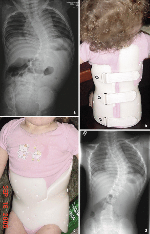

Fig. 28.6
EOS with tethered spinal cord. Scoliosis noted at the age of 6 months. No treatment was initiated and no MRI was done until the age of 2 years. In spite of detethering performed at the age of 2 years, scoliosis progressed at the age of 2 1/2 years (a) and bracing was initiated. Full-time custom-molded Boston brace is well-tolerated, asymmetric, and has large areas of relief opposite any area of pressure (b, c). At the age of 6 years, bracing continues with some progression of curve and mild thoracic deformity (d). Dual growing rods was planned, if worsening continued
Indications for orthotic treatment of older patients with idiopathic EOS are suggested by published results of brace treatment, likelihood of curve progression, and biomechanical curve simulations. This center has utilized a Cobb angle in excess of 20° as a lower threshold for orthotic treatment in idiopathic EOS curves. We agree with Winter [38] and urge that curves over 20° should be considered for brace treatment in EOS, assuming that the curve has been persistent or progressive and is in a region of the spine accessible to bracing. Biomechanical models [25, 39, 40] of scoliosis suggest that at approximately 25° of curvature, the load required to deform the spine diminishes significantly and that conversely, if the curve can be diminished to well under 25°, then vertebrae may be loaded much more symmetrically. Stokes et al. demonstrated asymmetric growth of rat tail vertebrae in response to asymmetric loading [26]. The rationale for early bracing of moderate curves (those in excess of 20°) is the assumption that by placing the growing spine under straighter mechanical load, there is some chance for spine remodeling toward symmetry, and progression is less likely during the preadolescent period of rapid growth. Sanders et al. [41] and many others have shown the early adolescent growth phase to be the period of greatest risk for progression of scoliosis, while Lonstein and Carlson [42] and Charles et al. [43] quantified the relationship between growth phase, curve magnitude, and the risk of progression. The goal of early bracing of moderate idiopathic EOS should be to enter the rapid preadolescent growth phase, when risk of progression is highest, with as little deformity as possible.
Early onset scoliosis associated with syringomyelia, Chiari malformation, or a tethered spinal cord should also be considered for brace treatment. Although surgical treatment of the Chiari malformation, syringomyelia, or tethered cord often results in improvement in the associated spinal deformity, the spinal deformity may continue to worsen if there is established deformity or persistent neuraxis abnormality [44]. Surgeons and families may falsely assume that by alleviating the presumed etiologic cause of the deformity, the deformity itself will probably resolve spontaneously as it frequently does if the deformity is mild. Many patients with established kyphotic deformity or scoliosis in excess of approximately 30° may develop progressive deformity during the rapid growth of preadolescence, even though the neuraxis abnormality has been treated. Although a trial of observation following decompression of the Chiari malformation or syringomyelia or tethered cord is appropriate, if the deformity persists as excessive kyphosis or greater than 20° of scoliosis, treatment as in idiopathic EOS should be instituted.
28.3.3 Contraindications
Contraindications to bracing include certain curve locations, very large curves, associated thoracic lordosis, advanced chest deformity, and some medical and psychological conditions. Multiple reports of brace treatment of adolescent idiopathic scoliosis note the poor results of bracing in upper thoracic curves, triple curves, and curves at the lumbo-sacral junction. Although the Milwaukee brace is felt to be the most appropriate brace for curves with apices above T6, reported results [45] are not encouraging, leading most practitioners to observe rather than treat the upper thoracic curves. Jarvis et al. [20], Lenke and Dobbs [21], and McMaster and Macnicole [18], in their series of idiopathic EOS, did not specifically note curves with predominantly high thoracic apices, suggesting that this is an uncommon curve pattern in this age group. Jarvis et al. [20] noted more success in EOS with single thoracic and thoracolumbar curves than with double major curves, mirroring our experience with adolescent idiopathic scoliosis and idiopathic EOS. Most curves with high thoracic apices are accompanied by secondary, less structural lower curves of lesser magnitude, which may be successfully treated by bracing. Surgical correction of the high thoracic curve followed by brace treatment of the lower curve(s) is also an option when the upper thoracic curve is rapidly progressive.
Thoracic hypokyphosis or thoracic lordosis is a nearly universal accompaniment of idiopathic thoracic curves, and is often cited as a contraindication to brace treatment, yet Mannherz et al. [22] found thoracic hypokyphosis in only 20 % of their series of idiopathic EOS curves. Although frequently mentioned [22, 23, 29, 45, 46], guidelines for the treatment of thoracic lordosis and hypokyphosis are not clear. Our practice has been to treat associated thoracic hypokyphosis with a brace modified to include posterior cephalad extensions of the brace intended to encourage thoracic kyphosis. For true thoracic lordosis (less than 0° of thoracic kyphosis), bracing may be counterproductive and produce more thoracic lordosis. Thoracic lordosis, in association with thoracic scoliosis, may however be the ideal indication for surgical guided-growth procedures such as anterior vertebral stapling, or tethering [47, 48].
Large curves (in excess of 60–90°) are rarely permanently stabilized by repetitive casting or bracing in EOS. Although bracing may be used for large curves to allow more growth before a planned surgical intervention, many large curves are best dealt with by surgical intervention such as dual growing rods. Moderately large curves in the adolescent may be successfully treated with bracing as demonstrated by Wiley et al. [49] and Katz and Durrani [50] and also in some EOS. In our Boston Brace series [29], none of the EOS patients, aged 4–10 years, with curves 40–49° at initiation of bracing needed surgery after bracing and follow-up. Our present practice is to attempt bracing in larger EOS curves between 40° and 60°, provided that there is acceptable chest deformity, but will switch to dual growing rods if the chest deformity worsens significantly.
Significant chest deformity [35, 51] often accompanies more severe EOS curves and may be a contraindication to bracing. Continued brace treatment may worsen the chest deformity while seemingly stabilizing the spine deformity. The more advanced the chest deformity is, the more likely that the patient will be left at the end of surgical treatment with a functionally significant thoracic deformity and increased risk of respiratory insufficiency as an adult [35, 37]. In most severe idiopathic EOS, final instrumentation and fusion is generally successful in achieving a balanced, stable, minimally deformed spine at the end of treatment. However, surgical treatment is rarely successful in restoring normal chest shape and normal chest compliance when there has already been a severe chest deformity. Therefore, chest deformity should not be allowed to worsen beyond a point at which it is irrevocable. Surgical treatment such as dual growing rods should be instituted earlier.
Stay updated, free articles. Join our Telegram channel

Full access? Get Clinical Tree


