Fig. 27.1
Lateral radiographs of the thoraco-lumbar spine demonstrating kyphosis progression in a 8 year old child. (a) The deformity measures only 30 degrees at the completion of chemotherapy. Over a period of 5 year follow-up, the deformity gradually worsens from 42° at 5 years (b) to 71° at the end of 10 years (c) Note that the T11 vertebra (arrow), which was uninvolved in the disease, is showing progressive destruction due to biomechanical influence during growth
27.2.2 Epidemiology
The global burden of tuberculosis still remains huge. The World Health Organization’s (WHO) Global Tuberculosis Report (2012) observes that there were an estimated 8.6 million new cases of tuberculosis and 1.3 million people died due to the disease [2]. The prevalence of tuberculosis is inversely related to the socioeconomic status of the society and the standard of public health. In developing countries, many children present late with significant deformity and neurological deficits. In developed countries, the diagnosis can be missed as it does not feature as a common diagnosis in the clinician’s mind.
In developing parts of the world with a high burden of pulmonary tuberculosis, the incidence of spinal infections is expected to be proportionately high, with India and China accounting for 26 % and 12 % of the global disease burden respectively in 2012 [2]. Though the exact incidence and prevalence of spinal tuberculosis in children are not known, the incidence of pediatric spinal tuberculosis is reported as 58 % of all spinal tuberculosis in Korea, 30 % of all patients treated for spinal tuberculosis in India, and 26 % in Hong Kong [3–5].
27.2.3 Microbiology and Pathophysiology
Tuberculosis is caused by a bacillus of the Mycobacterium tuberculosis complex. There are approximately 60 known species among the Mycobacterium genus but only a minority of these cause human tuberculosis (M. tuberculosis being the most common). Vertebral infection by the bacillus results from hematogenous dissemination from a primary focus elsewhere in the system, commonly the lungs and the kidneys. Spread of the organism can also occur through the lymphatics from viscera to the adjacent vertebral segments (e.g., pulmonary tuberculosis can spread to the thoracic spine).
Following the infection in the vertebral marrow, the inflammatory response is characterized by chronic accumulation of macrophages and monocytes. The tubercle bacilli are phagocytosed and their lipid is dispersed throughout the cytoplasm of macrophages, transforming the macrophages into epitheloid cells, which are characteristic of the tuberculous reaction. Another characteristic feature of tuberculous lesion is the presence of Langerhans giant cells, which are formed by the coalescence of a number of epitheloid cells. The typical histopathological lesion of tuberculosis is called the tubercle, which is formed by the conglomeration of macrophages, epitheloid cells, Langhans giant cells, lymphocytes, and inflammatory exudate. With progressive destruction, caseation necrosis occurs in the center of the tubercle. Adjacent tubercles then coalesce to form a large abscess and since it is a chronic infection, the acute features of inflammation like warmth and redness are absent (cold abscess).
27.2.3.1 Clinical Pathology
The most common pattern of tubercular spinal infection in adults is the “paradiscal” type, where the bacilli lodge in the sub-chondral marrow on either side of the disc as the disc is avascular. In children, the disc retains its blood supply till approximately 9 years of age and so the bacilli affect and destroy the vertebral body and disc simultaneously (“centrum” or “complete” type) (Fig. 27.2a–f). Due to the weaker immune response of the child and cartilaginous nature of the vertebral body, extensive vertebral destruction and exuberant abscess formation is more common in children [6]. The other types of spinal tuberculosis are the anterior type (abscess formation beneath the anterior longitudinal ligament), posterior type (isolated involvement of posterior elements), and the non-osseous type (extensive abscess formation with very little bony destruction).
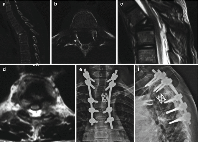

Fig. 27.2
Centrum (complete) type of tuberculosis in a 10-year-old child. Sagittal CT (a) and coronal CT (b) images show complete collapse of the T3 vertebra with involvement of the pedicles and lamina on the right side. Sagittal and axial MRI images (c, d) show complete collapse of the T3 vertebral body with prevertebral abscess formation. Unlike a classical adult paradiscal type of spinal tuberculosis, the centrum type is characterized by vertebral body destruction with intact adjacent disc spaces. The lesion has been treated by posterior stabilization, corpectomy, and reconstruction with cage as shown in the AP and lateral radiographs of the thoracic spine (e, f)
27.2.4 Clinical Presentation
Unlike pyogenic spondylitis, tuberculous lesions have a much more insidious onset and the clinical symptoms often develop over a period of 1–2 months. Back pain localized to the affected site and aggravated with spinal movements is the usual presenting feature. In the initial stages, the pain is due to inflammation, distension of the capsule by the abscess and pressure on neighboring structures. Later with development of instability, the pain can become quite severe. The affected child may need to support his trunk by placing the hands on the couch while sitting (Tripod sign) or hold the neck by the hands when the cervical spine is affected (Fig. 27.3a, b). Constitutional symptoms of malaise, loss of appetite and weight, evening rise of temperature and night sweats is also observed in up to 60 % of patients [7].
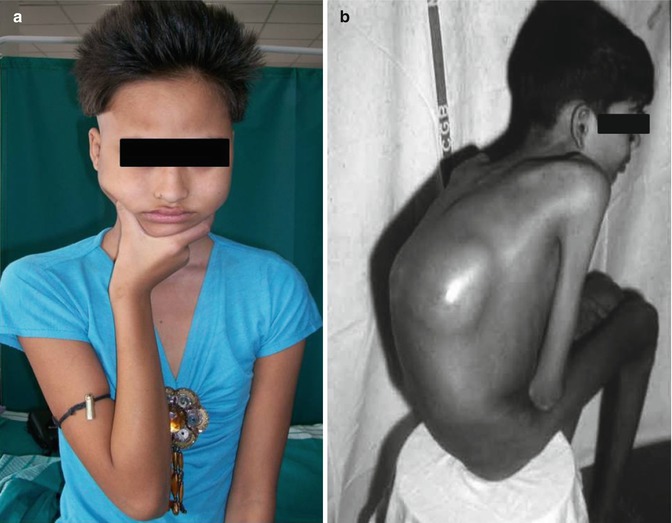

Fig. 27.3
(a) A 13-year-old girl with upper cervical tuberculosis and cervical instability holding her neck because of severe instability pain. (b) Another 9-year-old boy with prominent thoracolumbar kyphosis with a large lumbar abscess formation. Note that the patient is supporting his trunk with his elbows and the generalized wasting of the muscles. (b) (Courtesy: Prof. V.T. Ingalhalikar, India)
A paravertebral cold abscess is a diagnostic feature of spinal tuberculosis. It may be clinically evident, either in the paraspinal area or the abscess may tract distally along the perineural, perivascular, intermuscular, subpleural, subperitoneal, and natural areolar tissue spaces to present remotely away from the vertebral lesion. The common areas of presentation include the retropharyngeal abscess from a cervical lesion, a paravertebral abscesses in the thoracic spine tracking along the intercostal neurovascular bundle along the chest wall, and pre-sacral and pelvic retroperitoneal abscess from a lumbar lesion. A psoas abscess is common in thoracolumbar lesions below the diaphragmatic attachment to the spine. Psoas abscesses are pathognomonic of spinal tuberculosis and can present bilaterally. They can present externally in the inguinal region, at the Petit’s triangle (See Fig. 27.3b), in the ischiorectal fossa, or in the buttock under gluteus maximus (Fig. 27.4a–d).
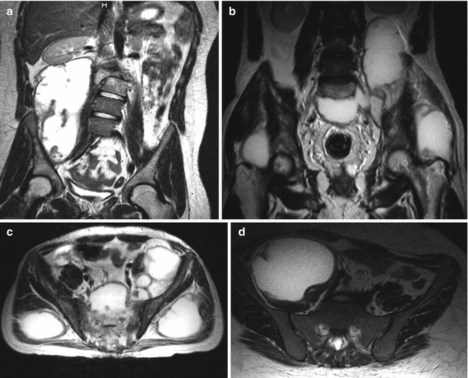

Fig. 27.4
Tubercular cold abscess can be present in multiple locations depending on the pathway of its spread across areolar spaces. In this 40-year-old patient, who presented with back and bilateral gluteal pain, extensive cold abscess formation was observed. Coronal T2 MRI sections of the lumbar spine (a, b) showing a right retroperitoneal psoas abscess (a), bilateral trochanteric abscesses (b). Axial T2 sections through the sacrum and pelvis shows multiple abscesses in iliac fossa (d), presacral and sub-gluteal regions (c), and large pre-sacral abscess (d)
Deeper abscesses are not clinically palpable but can cause pressure symptoms. A retropharyngeal abscess arising from cervical tuberculosis can produce dysphagia and dysphonia. In children with high thoracic spinal involvement with paraspinal abscess formation, the abscess in the prevertebral region may cause significant bronchial compression. These symptoms may simulate bronchial asthma as the dyspneic symptoms exacerbate when the patient lies down at night.
While most abscesses resolve gradually with chemotherapy treatment, they may also rupture with neglect leading to the formation of a sinus. The sinus may heal spontaneously with medical treatment after all the necrotic material is discharged or may persist if there is any residual infection or secondary pyogenic infection. The tubercular pus is white or light gray in color, watery, and has no specific smell unlike a pyogenic abscess.
27.2.4.1 Neurological Involvement
Neurological compromise occurs in up to 30–75 % of the patients with spinal tuberculosis [7–9]. Though children have more severe destruction, they also have a lesser incidence of neurological involvement, probably due to the relative larger canal diameter and more flexibility of the spine. Cervical tuberculous lesions manifest with quadriparesis but since the thoracic and thoracolumbar regions are commonly affected, lower limb weakness with bladder and bowel involvement is more common. Initial symptoms are in-coordination and clumsiness while walking which slowly progresses to paraplegia and loss of sphincter control.
Children can present with neurological involvement both in the active and healed phase of the disease. In active lesions, it is due to the result of direct compression of the spinal cord by an abscess, inflammatory granulation tissue, a dislodged sequestrum, or canal compromise due to instability. In the healed disease, it occurs after many years and is usually due to stretching of the cord over a bony ridge at the apex of the deformity.
27.2.4.2 Kyphotic Deformity
The etiology and progression of kyphosis is different in the active and healed phase of the disease. Tuberculosis affects and destroys the anterior structures of the vertebral column in more than 90 % of patients. Collapse of the vertebral body is evident as a localized kyphotic deformity. Involvement of two or three adjacent vertebral bodies manifests as a sharp, angular kyphosis called the gibbus. Chemotherapy will cure the disease but vertebral collapse will continue until the healthy vertebral bodies in the region of the kyphosis meet anteriorly and consolidate. The severity of collapse during the active phase is mainly influenced by the severity of vertebral destruction, the level of the lesion, and the age of the patient [10].
During the active phase, the deformity increases in proportion to the severity ranging about 25–35° for each vertebral body loss in thoracic and thoracolumbar lesions. The kyphotic collapse is less in lumbar lesions due to the lordotic nature of the lumbar spine, large size of the intervertebral discs, and the sagittal orientation of the facet joints which allows vertical subsidence. In the thoracic and the thoracolumbar regions, the deformity tends to be more severe due to the inherent kyphotic nature of the thoracic spine and the coronal orientation of the facets which lead to subluxation and kyphosis. Deformities in children less than 10 years of age have been observed to have greater deformity than those over 10 years of age due to soft vertebral bodies, weaker posterior stabilizing structures, and the secondary increase in deformity during the adolescent growth spurt [11, 12].
In a long-term follow-up of 15 years of 63 children, Rajasekaran reported three types of collapse and healing of the anterior column with different implications for deformity progression during the period of growth [10, 13, 14] (Fig. 27.5a–c). Type A healing was seen in minimal lesions and paradiscal type of involvement, where the facet joints were intact and there was large area of contact of vertebral bodies anteriorly. These patients showed minimal deformity in the active phase and frequently an improvement during the growth period (Fig. 27.6a–c). Type B healing was seen when the vertebral body loss is equivalent to the loss of one vertebral body. Here during the process of collapse, the facet joint at the level of destruction subluxed or completely dislocated. The superior vertebra rotates during the process of descent so that its antero-inferior margin comes into point contact with the superior surface of the inferior normal vertebra. This resulted in growth depression at the point of contact and the deformity could progress by up to further 30° during the growth period (Fig. 27.7a–c). Type C restabilization occurred when the vertebral body loss increases to more than “two.” The large anterior column defect necessitates the dislocation of two or more facet joints before anterior column restabilization can occur. The superior normal vertebra rotates 90° so that the anterior surface of superior vertebra comes into contact with the superior surface of the inferior vertebra. This was seen frequently in children less than 7 years of age with thoracolumbar disease. In children with multiple vertebral body destruction, a peculiar pattern of collapse is seen which has been termed as “Buckling Collapse” [14, 15]. Dislocation of facet joints occurs sequentially at multiple levels leading to a kyphosis of more than 120° and the entire spine is converted to two large compensatory curves. Many vertebral segments become horizontally oriented with stress shielding of their growth plates. Longitudinal overgrowth of the vertebral segments is noted leading to stretching of the spinal cord at the apex of the kyphosis with possible secondary late-onset paraplegia. Risk factors for “buckling collapse” included an age of less than 7 years at the time of the disease, thoracolumbar involvement, loss of more than two vertebral bodies, and presence of radiographic spine-at-risk signs (Fig. 27.8a–c).
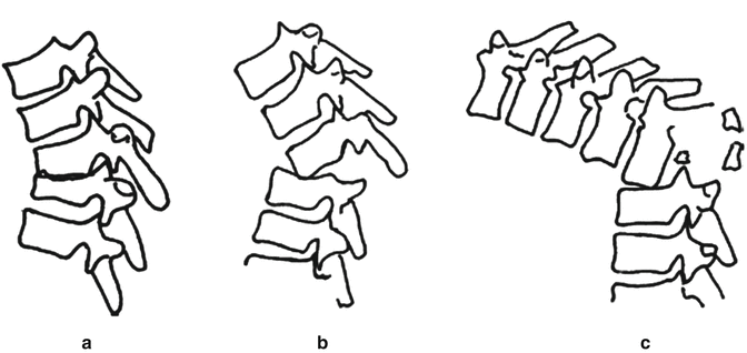
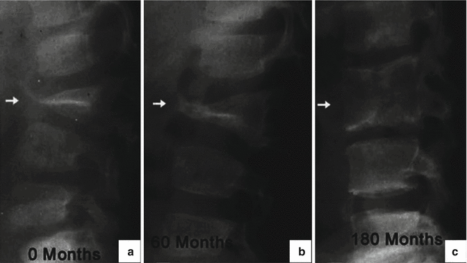
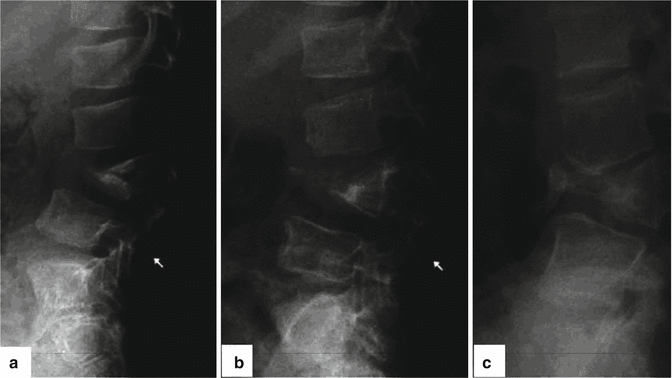
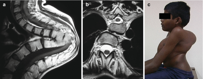

Fig. 27.5
Following destruction of anterior column, restabilization and healing occurs by one of the three methods. (a) In patients with minimally destroyed vertebrae with intact facet joints, restabilization occurs with wide contact area. (b) In patients with dislocation of single facet joint, restabilization occurred by point contact. (c) In patients with loss of two or three vertebrae, the facets dislocate at multiple levels and the superior segment rotates by 90° so that its anterior surface can rest on the superior surface of inferior vertebra

Fig. 27.6
Type A restabilization. In patients with partially destroyed vertebrae, restabilization occurs with wide contact area and the kyphosis gets corrected automatically or remains unchanged. (a) Lateral radiograph of thoracolumbar spine showing a healed tuberculous lesion at L2-3 which has healed into a triangle shaped fusion mass (arrow). The bony fusion has occurred over a wide contact area with intact facet joint. (b) During growth, the fusion mass shows “accelerated growth phenomenon” (arrow) and achieves spontaneous improvement in vertebral height at 60 months. (c) At 180 months, the fusion mass has become a large “single” vertebra (arrow) with two subjacent pedicles

Fig. 27.7
Type B restabilization. Type B healing is seen when the vertebral body loss is between 1 and 1.5. (a) Lateral radiograph of the lumbar spine shows significant destruction following L3-4 spondylodiscitis which has resulted in local kyphosis. (b, c) Lateral radiographs performed at 60 and 180 months show that the facet joint at the level of destruction is dislocated (white arrow) during the process of collapse and the superior L2 vertebra gradually rotates by 90 degrees so that its antero-inferior margin comes into point contact with the superior surface of the fusion mass

Fig. 27.8
Buckling collapse due to neglected tubercular kyphotic deformity in a child. The sagittal MRI of spine shows buckled spine with two long spinal segments lying on each other (a). The spinal cord is stretched and compressed at the apex of the kyphosis. The axial MRI image shows two vertebral segments at the same level straddled over each other because of buckling (b). Clinical picture of the same patient shows the shortened trunk because of buckling (c)
Unlike adults in whom the deformity is static after cure of the disease, post-tuberculous kyphosis in children is a dynamic deformity with variable progression during growth. Three different patterns of progression have been observed depending on the pattern of healing [10]. Type 1 progression, where worsening of deformity occurs during growth, is seen in 39 %. This increase can occur after a lag period of few years after the disease control. As a result, a severe increase in deformity may be missed if the child is not followed-up carefully till the completion of growth. Forty-four percent of children had a Type II progression where after an increase in deformity during the active phase, the deformity showed a progressive and spontaneous correction. This was mostly observed with Type A healing pattern and in children younger than 7 years. Type III progression, where there was no major change during growth, was seen in the remaining 17 % who either had a minimal disease or a lower lumbar lesion (Fig. 27.9a, b).


Fig. 27.9
Deformity progression in healed tuberculosis in adults and children. (a) In adults, the deformity remains the same during the healed phase. (b) In children, the deformity can either worsen (Type I), remain static (Type III), or improve (Type II) during the healed phase
Four radiological signs which indicate spinal instability have been identified by Rajasekaran to predict the risk of late and progressive development of deformity in childhood spinal tuberculosis [10, 13]. They basically indicate the presence of facet joint dislocation and disruption of the posterior arch. These signs are easy to identify in radiographs, appear early in the course of the disease and are useful to identify children at risk for progression so that surgical stabilization can be suitably advocated. These four “spine-at-risk” signs are: (1) dislocation of one or more facet joints in the lateral view, (2) retropulsion of the diseased vertebra, (3) lateral translation seen in antero-posterior view, and (4) the “Toppling Sign” (Fig. 27.10a–d).
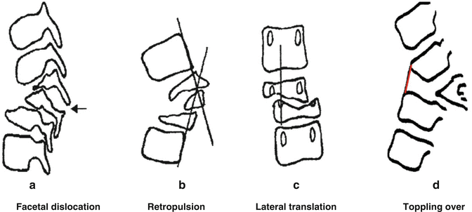

Fig. 27.10
Rajasekaran’s ‘Spine at risk’ radiological signs. (a) Separation of the facet joint. The facet joint dislocates at the level of the apex of the curve, causing instability and loss of alignment. In severe cases the separation can occur at two levels. (b) Posterior retropulsion. This is identified by drawing two lines along the posterior surface of the first upper and lower normal vertebrae. The diseased segments are found to be posterior to the intersection of the lines. (c) Lateral translation. This is confirmed when a vertical line drawn through the middle of the pedicle of the first lower normal vertebra does not touch the pedicle of the first upper normal vertebra. (d) Toppling sign. In the initial stages of collapse, a line drawn along the anterior surface of the first lower normal vertebra intersects the inferior surface of the first upper normal vertebra (red line). ‘Tilt’ or ‘toppling’ occurs when the line intersects higher than the middle of the anterior surface of the first normal upper vertebra
27.2.5 Diagnostic Investigations
Diagnosis may be difficult in children, especially in the early stages of the disease. Systemic symptoms of tuberculosis include malaise, easy fatigability, weight and appetite loss, and sometimes low grade fever. Clinical signs of frank sepsis are uncommon.
27.2.5.1 Laboratory Investigations
Anemia and elevated erythrocyte sedimentation rate (ESR) are the two common abnormalities noted in blood investigations. ESR may be markedly elevated (>70 mm/h) and serial ESR measurements are helpful in assessing the response to treatment. ESR and low hemoglobin levels however lack specificity [16]. A positive Mantoux (tuberculin skin) test merely indicates cell-mediated immune response due to a previous tuberculous infection and in endemic regions, the test can be positive even in patients without active tuberculosis. Its diagnostic value is useful only in regions where tuberculosis is rare. Polymerase chain reaction (PCR) analysis from infected tissue is considered very sensitive and specific for the diagnosis of spinal tuberculosis [17].
27.2.5.2 Bacterial Cultures
Bacterial culture of the infected tissue is useful to confirm the diagnosis and to acquire antibiotic sensitivities to guide therapy. Since spinal tuberculous infection is paucibacillary (less bacilli in infected tissues), it is essential to culture material from deep structures such as bone and abscess walls. Culture media such as BACTEC™ (Becton-Dickinson and Co., USA) now are the standard culture media [18]. An important advantage is that they allow drug susceptibility assessment. This helps in identifying drug resistant strains and start early alternate second-line medications.
Histopathology and Microbiology
The confirmation of tuberculosis infection is through identification of bacillus in the tissue or by histological confirmation of typical tubercles in the infected tissue. The typical histopathological findings are large caseating necrotizing granulomatous lesions with epitheloid and multinucleated giant cells with lymphocytic infiltration [19].
27.2.5.3 Imaging Studies
Earliest features observed on plain radiographs are vertebral osteoporosis, narrowing of the disc space and indistinct paradiscal margin of vertebral bodies. With progress of the disease, destruction is associated with vertebral collapse, kyphosis, and sagittal or coronal instability. In the cervical spine, the prevertebral soft tissue shadow can be enlarged due to distension of the abscess in the retropharyngeal region. In the thoracic spine, the cold abscess is visible on antero-posterior plain radiographs as a fusiform or globular radiodense shadow (bird’s nest appearance) (Fig. 27.11a–e). In children, attention should be paid toward the “spine-at-risk signs” as it indicates chances of deformity progression.
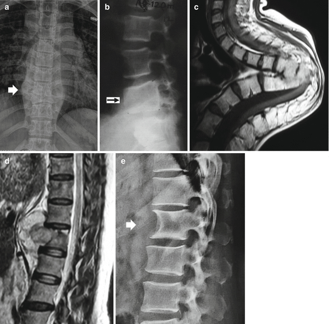

Fig. 27.11


Radiological appearances in spinal tuberculosis. (a) Bird’s nest appearance: In the anteroposterior radiograph of the spine, the paravertebral abscess formation is seen as a fusiform shaped radiodense shadow (thick white arrow). (b) Vertebra within vertebra appearance – Healing of a paradiscal type of tuberculosis results in fusion of two adjacent vertebral bodies, which is seen as a single vertebra with two subjacent pedicles (black arrow). (c) Horizontalisation of vertebral body in buckling collapse – Chronic buckling collapse can lead to increase in the supero-inferior height of the horizontally placed vertebral bodies. (d, e) Aneursymal phenomenon – Sagittal MR image shows prevertebral abscess formation under the anterior longitudinal ligament. Such longstanding abscesses can cause erosion of the anterior surface of the vertebral body (E) (thick white arrow) similar to an aortic aneurysm
Stay updated, free articles. Join our Telegram channel

Full access? Get Clinical Tree


