Fig. 3.1
Comparison between 18F-FDG PET/CT and 68Ga-DOTANOC PET/CT in a patient with a nodule in the pancreatic tail incidentally detected at CT. 18F-FDG PET/CT showed no 18F-FDG uptake in the pancreatic lesion (a, b) (arrow), while 68Ga-DOTANOC PET/CT showed intense focal tracer uptake (c, d) (arrow), thus suggesting the presence of a pancreatic NEN. The patient underwent distal pancreasectomy; surprisingly histology showed the presence of an intrapancreatic spleen. (e) Pancreatic tissue showing a peripheral (left upper corner) well-demarcated nodule approximately 1 cm in size corresponding to a benign intrapancreatic accessory spleen (f). H&E (e, f) (Courtesy of Dr. Frediano Inzani, Institute of Pathology, Università Cattolica del Sacro Cuore, Rome, Italy)
18 F–DOPA. Dihydroxyphenylalanine (DOPA) is an amino acid intermediate in the catecholamine synthesis pathway, which is taken up into the cells via the large neutral amino acid transporter and converted to dopamine by aromatic amino acid decarboxylase (AADC) [23]; via the vesicular monoamine transporters, DOPA is stored in the secretory granules where enzymatic degradation is prevented [24]. AADC is usually upregulated in neuroendocrine tumor cells, especially pheochromocytomas/paragangliomas and gastroenteric NENs with the exception of pancreatic islet cell tumors, where lower AADC levels have been reported [24]. For imaging purposes, DOPA can be labeled with both 11C and 18F; however, 11C-DOPA is not widely used because of limited availability of 11C (physical half-life of 20 min with need of an on-site cyclotron for production) and high costs. 18F-DOPA has been registered in France in 2006 and is commercially available in Europe but not in the USA. A potential limitation of 18F-DOPA is the physiologic uptake in the abdomen (liver, gallbladder, biliary tract, pancreas, and duodenum), which might mask tumors in this site [2]. Premedication with carbidopa, an inhibitor of the peripheral AADC, can improve image quality by decreasing background activity [23]. 18F-DOPA PET/CT performs well in patients with functioning carcinoid tumors, with lower sensitivity for pancreatic islet cell tumors due to the physiologic pancreatic uptake [25].
11 C–5-HTP. 11C-5-hydroxytryptophan (11C-5-HTP) is a serotonin precursor and DOPA decarboxylase substrate, which is specifically and irreversibly trapped by serotonin-producing tumors. Unfortunately, 11C-5-HTP is produced only in centers with a cyclotron on site and at high costs; so, its use is essentially limited to clinical trials in specialized centers [26]. As for 18F-DOPA PET, carbidopa premedication increases tumor-to-background ratio so improving image interpretation [27]. 11C-5-HTP PET seems to be useful in many tumor entities of the heterogeneous NEN group, especially in detecting small pancreatic neoplasms [28].
18 F–FDG. 18F-Fluorodeoxyglucose (FDG) is a glucose analog and the most used PET tracer in oncology with very high sensitivity in many types of tumors, especially the rapid-growing and aggressive ones. FDG uptake in neoplastic cells reflects the highest glucose metabolism which is linked to the cellular proliferative activity [2]. Procedure guidelines for PET and PET/CT tumor imaging with 18F-FDG have been published in Europe, the USA, and Japan [29–31]. Pitfalls in the interpretation of FDG images may be caused by the physiologic activity in the brain, vocal cords, esophagus, heart, stomach, bowel, bladder, and brown fat. An increased FDG uptake can be seen also in infections and other inflammatory processes [2, 30]. The usefulness of FGD PET/CT in the diagnosis of NEN is strictly related to the grade of differentiation, as high 18F-FDG uptake is usually associated with more aggressive GEP-NENs and a less favorable prognosis [28].
3.3 New Tracers for SPECT and PET
Besides radiolabeled somatostatin analogs, the use of radiolabeled somatostatin antagonists represents a highly promising strategy for both SPECT and PET imaging [1, 32]. Preliminary experiences showed that the radiolabeled antagonist 111In-DOTA-BASS may be preferable to agonists because of a higher and longer retention in tumors and better imaging results [33]; so, a greater benefit on radioreceptor therapy is expected [34]. Among new SPECT tracers, specific radioligands for glucagon-like peptide-1 (GLP-1) receptors have been recently developed and evaluated in animal models and in humans [35, 36]. GLP-1 is an intestinal hormone that stimulates insulin secretion through receptors expressed in islet cells. GLP-1 receptors are greatly expressed in insulinomas, and recent studies demonstrated that an 111In-labeled GLP-1 analog (111In-exendin-4) can detect occult insulinomas, which were not localized by other imaging modalities [35, 36]. 68Ga-exendin-4 has been recently applied for PET/CT imaging [37].
3.4 Clinical Applications
Many different radiopharmaceuticals are available for imaging of pancreatic neuroendocrine neoplasms (as other GEP-NENs) based on their physiopathology, which is characterized by different metabolic pathways and different receptor expression.
SRS with 111In-octreotide still today is a widely used functional imaging modality for GEP tumors, due to its worldwide availability together with good diagnostic sensitivity, which is ≥80 % in various studies with the only exception of insulinomas due to the low density of sstr2 in these tumors and its ability in changing patient management in approximately 30 % of cases [2]. 99mTc-labeled somatostatin analogs (99mTc-HYNIC-TOC and 99mTc-HYNIC-TATE) have been proposed as promising candidates for an alternative to 111In-DTPA-octreotide for SRS [6]. The main limitations of SRS are its relatively low spatial resolution (about 1 cm) and the lack of a precise localization of the neoplastic lesions when a SPECT/CT device is not available.
The initial clinical applications of 68Ga-DOTA-peptides have shown several advantages over 111In-DTPA-octreotide [38–40]: higher diagnostic sensitivity (>90 %), especially in detecting small primary tumors and metastatic lesions, due to the higher spatial resolution (4–6 mm) and higher count rate sensitivity of PET devices; quantification of the uptake by the standardized uptake value (SUV); lower radiation exposure; single-day procedure (versus 2–3 days for SRS if a hybrid machine is not available), with better patient comfort and faster reporting; and lower costs [40, 41]. Also the superiority of 68Ga-DOTATATE over 99mTc-HYNIC-TOC has been recently demonstrated [42]. 68Ga-DOTA-peptide PET/CT has shown to be the most sensitive procedure for the detection of unknown primary NENs; among the detected primary tumors, a significant number is located in the pancreas (46 % in the experience by Prasad et al.) [43]. In the presence of a pancreatic mass, a positive scan suggests the presence of a well-differentiated NEN (Fig. 3.2). On the contrary, a negative scan suggests an adenocarcinoma or a poorly differentiated neuroendocrine neoplasm; in these cases, an intense 18F-FDG uptake is frequently observed. In any case, preoperative cytologic/histologic confirmation is needed. In addition, 68Ga-DOTA-peptide PET/CT can affect either stage or therapy in approximately 50 % of patients with NEN [44] and plays a distinctive role in guiding the selection of therapy with cold or radiolabeled somatostatin analogs.
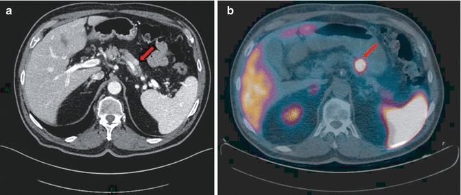
Fig. 3.2
(a) Axial contrast-enhanced CT image obtained in the arterial phase showing a 1 cm homogeneous hyperenhancing mass at the junction of the pancreatic body and tail (arrow). (b) Axial fused 68Ga-DOTANOC PET/CT images demonstrating intense and focal tracer uptake in the same pancreatic nodule, leading to the diagnosis of neoplastic disease characterized by elevated expression of somatostatin receptors (arrow)
18F-DOPA is particularly suited to visualize carcinoid tumors with elevated serotonin levels [2, 45] and is the imaging technique of choice in children with congenital hyperinsulinism to distinguish focal from diffuse forms [46, 47]. On the contrary, controversial results have been reported in localizing neuroendocrine pancreatic tumors in adults [48–50]. Due to the physiologic pancreatic uptake, false-negative findings with 18F-DOPA PET have been reported both in functioning and nonfunctioning neuroendocrine pancreatic tumors in adults as well as in patients with pancreatic islet cell tumors who had been pretreated with carbidopa, suggesting that carbidopa premedication should be avoided when such tumors are suspected [48]. Recent observations seem to reassess the value of 18F-DOPA PET/CT after carbidopa premedication in insulin-secreting tumors in adults [51]. Abnormal 18F-DOPA uptake in a pancreatic tumor not of neuroendocrine origin (i.e., solid pseudopapillary tumor) has been recently reported, doubting the specificity of this tracer [52].
The uptake of 11C-5-HTP in NENs is based on the same mechanism as for 18F-DOPA; however, few data are available about the use of 11C-5-HTP due to its complex production and limited availability. 11C-5-HTP can be used both in patients with increased serotonin synthesis, i.e., with carcinoid tumors, and patients without elevation of urinary 5-HIAA, i.e., with neuroendocrine pancreatic neoplasms [53]. In comparative studies, 11C-5-HTP PET showed the highest sensitivity (96 %) for the detection of pancreatic NENs as compared with CT, SRS, and 18F-DOPA PET [28]. False-negative results may occur in poorly differentiated neoplasms [48, 53].
18F-FDG frequently fails to visualize tumors with a low proliferation rate, i.e., well-differentiated NENs (G1), whereas it is recommended for identification and assessment of poorly differentiated (G3) neuroendocrine carcinomas [54, 55]. In patients with NEN, the so-called “flip flop” pattern of uptake may be observed, i.e., 68Ga-peptide positive and 18F-FDG negative in well-differentiated tumors and vice versa in poorly differentiated carcinomas [2] (Fig. 3.3). However, 18F-FDG positivity has been reported also in a substantial percentage of G1 and G2 NENs, where it offers important prognostic information, predicting early progression and influencing the therapeutic management [56]. Recently, a metabolic grading system based on the tumor-to-liver ratio of 18F-FDG uptake has been assessed in patients with inoperable multifocal gastroenteropancreatic NEN [57].
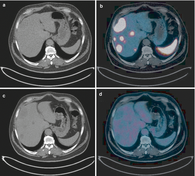
Fig. 3.3
Comparison between 68Ga-DOTANOC PET/CT and 18F-FDG PET/CT in a patient with liver metastases from previously removed well-differentiated pancreatic neuroendocrine tumor (G1). The images show multiple liver metastatic lesions characterized by intense 68Ga-DOTANOC uptake (a, b); the same lesions showed no FDG uptake (c, d), confirming the biological behavior of a G1 NEN
Considering the wide availability of radiopharmaceuticals that target endocrine tumor cells, how to choose the most appropriate one in the clinical practice for imaging endocrine pancreatic neoplasms? The choice depends on several factors including:
The biologic characteristics of the tumor, i.e., the metabolic activity as well as receptor subtype and specific amine profile, the functional activity, and the histologic grading (Ki-67)
The clinical information needed, i.e., diagnostic or prognostic
The diagnostic efficacy of each tracer in the various clinical situations, as well as the impact on patient management and on therapeutic decision
Practical issues, such as availability and costs as well as local expertise
Therefore, the preferential use of a given tracer should be defined in the specific clinical context (Table 3.1). When the diagnostic information is needed, i.e., for lesion identification and localization (including unknown primary) and disease staging and restaging after therapy, the choice should be primarily guided by tumor differentiation and grading. In well-differentiated pancreatic neuroendocrine tumors (G1 and low G2), functional imaging with radiolabeled peptides should be performed. In this context, 68Ga-DOTA-peptide PET/CT is currently regarded as the nuclear medicine technique of choice; where available, it has replaced OctreoScan®, which maintains a role just for evaluating the receptor status prior to therapy with cold or radiolabeled somatostatin analogs (90Y- or 177Lu-DOTA-peptides) [58]. In fact, PET/CT with 68Ga-DOTA-peptides provides the same information of SRS with a higher diagnostic accuracy and a lower patient discomfort.
Table 3.1
Choice of the radiopharmaceutical for imaging pancreatic neuroendocrine neoplasms
111In-DTPA-octreotide | 68Ga-DOTA-peptides | 18F-FDG | 18F-DOPA | |
|---|---|---|---|---|
Tumor biologic features | SMS receptor status | SMS receptor status | Glucose metabolism | Amine metabolism |
Diagnostic information needed (unknown primary, staging and restaging after therapy) | Obsolete | In well/moderately differentiated NENs (G1, low G2) | In poorly differentiated NENs (high G2, G3) | In well/moderately differentiated NENs (G1, low G2) |
Prognostic information needed | – | – | In G1, G2, G3 NENs | – |
Corresponding therapeutic information | Receptor status prior to therapy with cold or radiolabeled SMS analogs | Receptor status prior to therapy with cold or radiolabeled SMS analogs | – | – |
Practical issues | Registered, commercially available | Not registered on-site production
Related posts: Endocrinological Approach to the Diagnosis of Pancreatic Neuroendocrine Neoplasms
Molecular Pathology of Pancreatic Neuroendocrine Neoplasms
Immunohistochemical Approach to the Diagnosis and Prognostic Evaluation of Pancreatic Neuroendocrine Neoplasms
Glucagonoma
Mixed Adenoneuroendocrine Carcinoma of the Pancreas
ACTH-Producing Tumor Endocrinological Approach to the Diagnosis of Pancreatic Neuroendocrine Neoplasms
Molecular Pathology of Pancreatic Neuroendocrine Neoplasms
Immunohistochemical Approach to the Diagnosis and Prognostic Evaluation of Pancreatic Neuroendocrine Neoplasms
Glucagonoma
Mixed Adenoneuroendocrine Carcinoma of the Pancreas
ACTH-Producing Tumor
Stay updated, free articles. Join our Telegram channel
Full access? Get Clinical Tree
 Get Clinical Tree app for offline access

|