Vascular Dementias


Vascular Dementias


Nikolaos Scarmeas


INTRODUCTION

During much of the 20th century, it was assumed that most dementia of the elderly was vascular in origin, an assumption challenged during the last few decades after the recognition and importance of Alzheimer-type pathologic changes. Nevertheless, with wider use of modern sophisticated neuroimaging techniques and accumulation of more knowledge on cerebral microinfarcts, interest in the role of vascular disease on cognitive decline reemerged and the clinical importance of vascular lesions in cognitive performance has been a new focus of attention lately.

Brain injury caused by stroke can undoubtedly contribute to dementia and intellectual impairment. Vascular dementia is a progressive cognitive dysfunction caused by stroke, often ischemic, but also hemorrhagic cerebrovascular disease as well as ischemic white matter disease or sequelae of hypotension or hypoxia.

Diagnosis of vascular dementia has been controversial, and its definition has been unsettled despite numerous attempts at clarification. Unlike other neurodegenerative dementias, there are no specific pathologic criteria. Cerebrovascular disease is itself a heterogeneous disorder, with a variety of pathophysiologic mechanisms and clinical manifestations, and correspondingly, vascular dementia manifests also as a heterogeneous syndrome. Vascular dementia can result from a variety of mechanisms (Table 53.1 and Figs. 53.1, 53.2, 53.3).


TABLE 53.1 Proposed Types of Vascular Dementia Based on Localization and Pathophysiologic Mechanism


Vascular Dementia Subtype


Comments/Additional Information for Each Subtype, Including Localization, and Mechanisms


Critically located (strategic “single”) infarcts


Bilateral frontal infarctions from anterior cerebral artery occlusions


Bilateral thalamic infarction from posterior cerebral artery occlusions


Middle cerebral artery occlusions leading to frontal/parietal infarcts, etc.


Multiple ischemic infarcts affecting large vessels


Not necessarily in critical locations but leading to significant parenchymal volume loss


Small-vessel disease


Multiple lacunar infarcts in the basal ganglia


Multiple lacunar infarcts in subcortical or periventricular white matter leading to a more widespread, patchy pattern often described as leukoaraiosis in brain imaging studies (Binswanger disease)


Hemorrhagic infarcts


Intraparenchymal hemorrhages due to chronic vascular damage from hypertension


Amyloid angiopathy


Intracerebral or subdural hematomas or subarachnoid hemorrhage


Hypoperfusion


Due to systemic causes such as heart failure, hypotension, bypass surgery, cardiac arrest, medications, or other systemic conditions


Other mechanisms


Combinations of the above


Cerebral autosomal dominant arteriopathy with subcortical infarcts and leukoencephalopathy (CADASIL), an autosomal dominant form of cerebrovascular dementia (and also migraines and psychiatric symptoms) related to mutations in the notch3 gene on chromosome 19


Mitochondrial encephalomyopathy with lactic acidosis and stroke-like episodes (MELAS)


There are many criteria for the clinical diagnosis of “vascular dementia,” some not adequately validated, many inconsistently applied and in general lacking satisfactorily high sensitivity and specificity. In terms of terminology, the concept of “vascular cognitive impairment” has been also recently proposed; a more general term referring to “cognitive impairment that is caused by or associated with vascular factors” and includes severity stages corresponding to either dementia or mild cognitive impairment.

Overall, as a practical rule, a diagnosis of vascular dementia requires (1) clinically (symptoms/signs) and radiologically ascertained strokes (see Figs. 53.1, 53.2, 53.3), (2) dementia, and (3) a clear temporal relationship between stroke(s) and dementia.


EPIDEMIOLOGY

As many as 15% to 20% of patients with acute ischemic stroke older than age 60 years have dementia at the time of the stroke,
and 5% per year become demented thereafter. On a clinical basis, vascular dementia has been long considered as the second (after Alzheimer disease) or even the third (after Alzheimer and dementia with Lewy bodies) most frequent cause of dementia. Considering pathologic series data and related clinical-pathologic correlations makes the issue quite more complex.


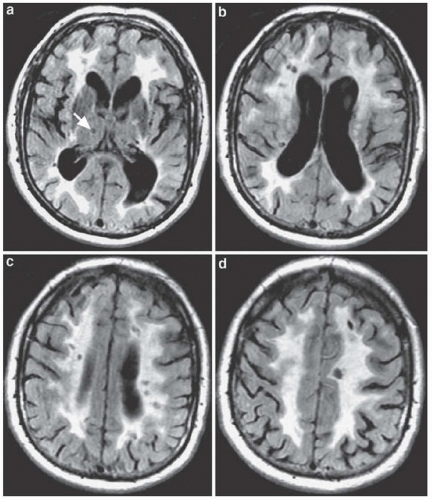
FIGURE 53.1 Binswanger disease in an 85-year-old man with dementia. A-D: Axial fluid-attenuated inversion recovery magnetic resonance images show extensive symmetric hyperintensity involving periventricular and lobar white matter. These lesions have a rather sharp outer border and show sparing of the U-fibers. This diffuse involvement was considered more than 25% of the total white matter. There are also a lacunar right thalamic infarct (arrow), several bilateral hypointense lesions within the periventricular hyperintensity representing lacunar infarcts and an important global cerebral atrophy. (From Guermazi A, Miaux Y, Rovira-Cañellas A, et al. Neuroradiological findings in vascular dementia. Neuroradiology. 2007;49[1]:1-22.)

Although the vast majority of elderly demented patients have Alzheimer-type pathologic changes (70% to 80%), approximately half of them have coexisting cerebrovascular infarctions too, whereas only about 30% have “pure” Alzheimer only pathology. Therefore, the majority of demented subjects have mixed Alzheimer and vascular pathologic changes. Adding to the argument of higher contribution rates of vascular dementia is the fact that most pathologic studies consider only larger infarcts, whereas microinfarcts (if time and resources permit their identification) are extremely common and diffuse in the brain. Finally, many white matter abnormalities visualized on brain magnetic resonance imaging are often undetectable by
standard neuropathologic examinations performed on autopsy, suggesting limited sensitivity of neuropathology for detection of cerebrovascular damage.


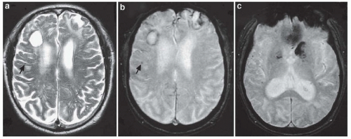
FIGURE 53.2 Amyloid angiopathy in a 66-year-old man with dementia. A: Axial spin-echo T2-weighted MR image shows bilateral large frontal hemorrhages surrounded by a rim of hypointensity due to hemosiderin deposition. There is also another right frontal punctate hypointense lesion (arrows), best seen on B, the axial gradient echo T2-weighted MR image. C: Axial gradient echo T2-weighted MR image caudal to B shows additional hypointensities bilaterally in the basal ganglia and the right parietal lobe (From Guermazi A, Miaux Y, Rovira-Cañellas A, et al. Neuroradiological findings in vascular dementia. Neuroradiology. 2007;49[1]:1-22.)

On the counterargument, the clinical salience of such microinfarcts on cognitive function has not been convincingly demonstrated. Additionally, dementia due to infarcts solely without significant pathologic contributions of the Alzheimer or Lewy body type (i.e., pure vascular dementia) is strikingly uncommon with estimated rates between 2% and 10% of dementias (not more than 5% of dementias in the United States).


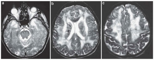
FIGURE 53.3 CADASIL in a 57-year-old man with cognitive impairment. A: Axial spin-echo T2-weighted MR images show an extensive white matter abnormalities with characteristic symmetric involvement of the anterior part of the temporal lobes and (B) external and extreme capsules without involvement of the internal capsules. They also show (C) extensive white matter changes in the centrum ovale, extending toward the cortex in some places. There are also two small lacunar infarcts in the right basal ganglia (arrowheads). (From Guermazi A, Miaux Y, Rovira-Cañellas A, et al. Neuroradiological findings in vascular dementia. Neuroradiology. 2007;49[1]:1-22.)

Only gold members can continue reading. Log In or Register to continue

Stay updated, free articles. Join our Telegram channel

Jul 27, 2016 | Posted by in NEUROLOGY | Comments Off on Vascular Dementias

Full access? Get Clinical Tree

Get Clinical Tree app for offline access