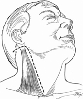
22 Eli M. Baron, Alexander R. Vaccaro, and Peter M. Fleischut The lateral retropharyngeal approach to the upper cervical spine was developed by Kelly and Whitesides as an alternative approach to the anterior cervical spine, avoiding the complexities of anterior extrapharyngeal approaches, which require an approach medial to the carotid sheath or dislocation of the mandible. This approach is actually done posterior to the carotid sheath, avoiding branches of the carotid artery and the facial nerve. Additionally, the approach allows access from C1 to T1 and avoids the potentially higher morbidity of a transoral/transpalatal approach. To perform a stand-alone C1-C2 anterior arthrodesis via a bilateral lateral approach. Instability at C1-C2 requiring anterior fixation or when an anterior approach is required for a diagnosis. The approach is advantageous over a transoral approach where fixation is necessary for instability and a posterior approach is contraindicated (presence of posterior infection or incompetent posterior elements). It also may be useful as a salvage technique following failed posterior arthrodesis. The patient is placed in the supine position. Fiberoptic nasotracheal intubation is preferred in cases of significant instability. Intraoperative neurophysiologic monitoring, including somatosensory evoked potentials and transcranial motor evoked potentials, if available, is used during positioning and throughout the procedure. Dental occlusion should be maintained to keep the angle of the mandible from limiting the area of dissection. Biplanar fluoroscopy or frameless stereotaxy may be useful during guidewire insertion, drilling, and screw placement to ensure proper instrumentation placement while limiting the risk of neurovascular injury. For positioning, if not contraindicated, the neck should be rotated to the opposite side and extended as much as possible. The ear lobe can be sewn anteriorly to the cheek to facilitate exposure of the field. Postoperative prophylactic tracheostomy should be considered in cases where there is significant retropharyngeal dissection. Performance of the tracheostomy after the procedure is usually more convenient. Preoperative imaging and planning are essential prior to performing the procedure. This includes computed tomography (CT) imaging for studying bony anatomy and estimating screw length. CT angiography and magnetic resonance angiography (MRA) are useful noninvasive modalities for assessing the position and patency of the vertebral arteries. A hockey-stick incision is made from the tip of the mastoid process and taken distally along the sternocleidomastoid muscle (Fig. 22.1). The greater auricular nerve is identified as it crosses the sternocleidomastoid muscle and dissected proximally and distally to increase its laxity, facilitating retraction. If needed, it may be divided with a resultant small sensory deficit around the ear. The external jugular vein is also ligated and divided.
Anterior C1, C2 Arthrodesis: Lateral Approach of Barbour and Whitesides
Description
Expectations
Indications
Contraindications
Special Instructions, Position, and Anesthesia
Tips, Pearls, and Lessons Learned
Difficulties Encountered
Key Procedural Steps
Related posts:
 Anterior Thoracic and Thoracolumbar Plating Techniques
 Anterior Cervical Corpectomy
 Posterior Spinal Anchor Strategy Placement and Rod Reduction Techniques: Posterior (Rotation vs. In-Situ Translation)
 Iliosacral Screw Fixation Techniques
 Open Lumbar Microscopic Diskectomy
 Spondylolysis Repair (Pars Interarticularis Repair)
Stay updated, free articles. Join our Telegram channel

Full access? Get Clinical Tree


