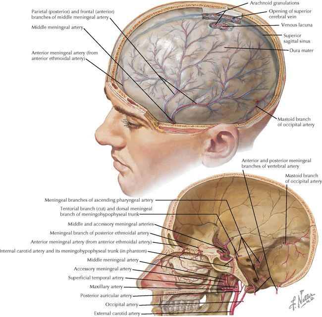
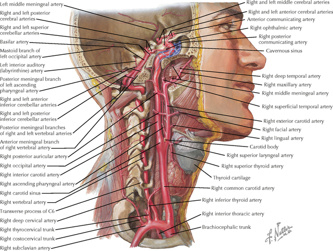
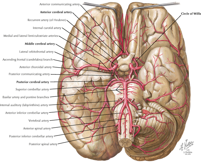
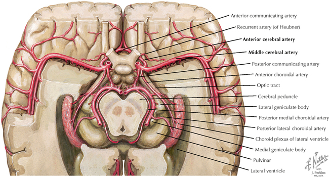
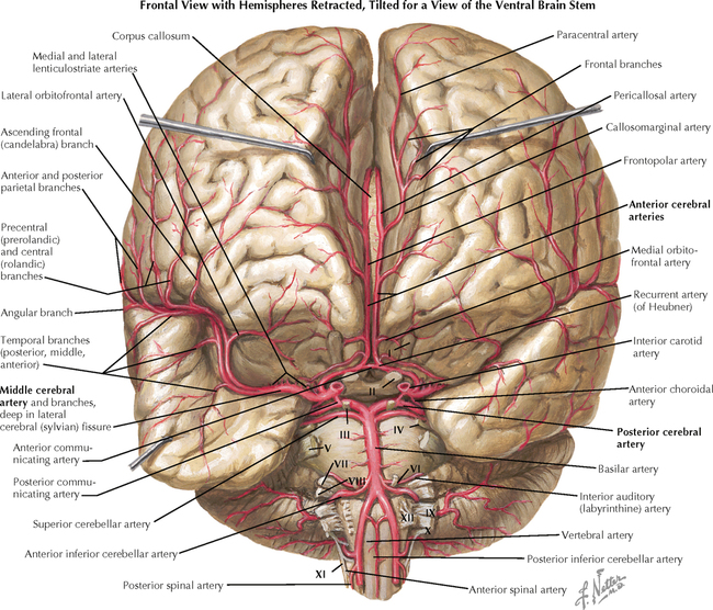
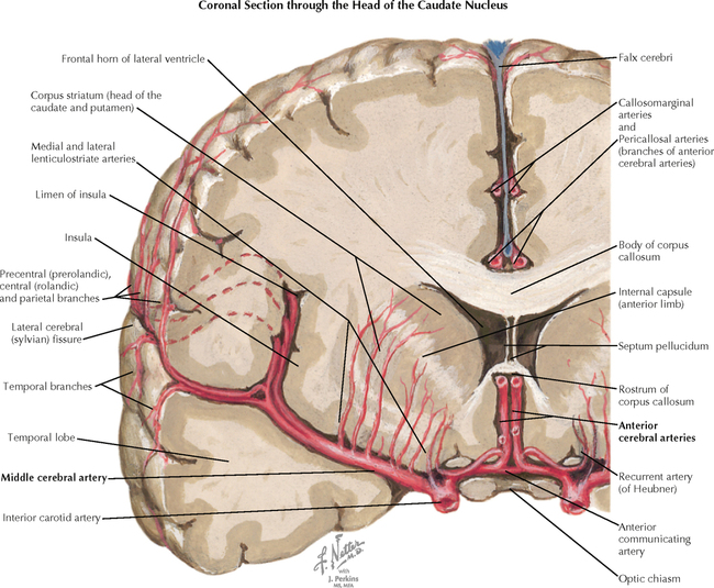
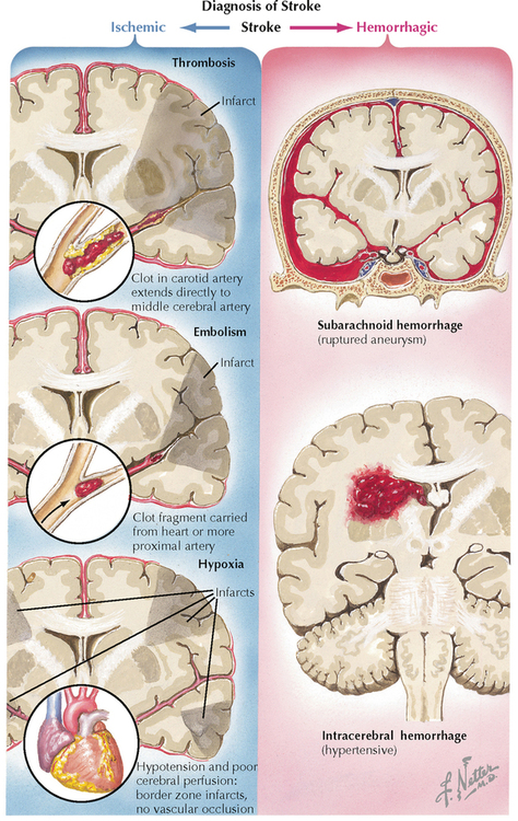
7 VASCULATURE Arterial System 7.1. Meningeal Arteries: Relationship to Skull and Dura 7.2. Arterial Supply to the Brain and Meninges 7.3. Internal Carotid and Ophthalmic Artery Course 7.4. Arterial Distribution to the Brain: Basal View 7.5. Arterial Distribution to the Brain: Cutaway Basal View Showing the Circle of Willis 7.6. Arterial Distribution to the Brain: Frontal View with Hemispheres Retracted 7.7. Arterial Distribution to the Brain: Coronal Forebrain Section 7.8. Types of Strokes 7.9. Schematic of Arteries to the Brain 7.10. Circle of Willis: Schematic Illustration and Vessels in Situ 7.11. Arterial Distribution to the Brain: Lateral and Medial Views 7.12. Color Illustration of Territories of the Cerebral Arteries 7.13. Magnetic Resonance Angiography: Frontal and Lateral Views 7.14. Angiographic Anatomy of the Internal Carotid Circulation 7.15. Vertebrobasilar Arterial System 7.16. Angiographic Anatomy of the Vertebrobasilar System 7.17. Vascular Supply to the Hypothalamus and the Pituitary Gland 7.18. Arterial Blood Supply to the Spinal Cord: Longitudinal View 7.19. Anterior and Posterior Spinal Arteries and Their Distribution 7.20. Arterial Supply to the Spinal Cord: Cross Sectional View Venous System 7.21. Meninges and Superficial Cerebral Veins 7.22. Veins: Superficial Cerebral, Meningeal, Diploic, and Emissary 7.23. Venous Sinuses 7.24. Deep Venous Drainage of the Brain 7.25. Deep Venous Drainage of the Brain: Relationship to the Ventricles 7.26. Carotid Venograms: Venous Phase 7.27. Magnetic Resonance Venography: Coronal and Sagittal Views 7.28. Venous Drainage of the Brain Stem and the Cerebellum 7.29. Venous Drainage of the Spinal Cord ARTERIAL SYSTEM 7.1 MENINGEAL ARTERIES: RELATIONSHIP TO SKULL AND DURA Meningeal arteries are found in the outer portion of the dura; they supply it with blood. They also help to supply blood to adjacent skull and have some anastomoses with cerebral arteries. The skull has grooves, or sulci, for the meningeal vessels. This relationship reflects an important functional consequence of skull fractures. Fractures can rip a meningeal artery (usually the middle meningeal artery) and allow arterial blood to accumulate above the dura. Such an epidural hematoma is a space-occupying mass and can produce increased intracranial pressure and risk for herniation of the brain, particularly across the free edge of the tentorium cerebelli. Even very fine fractures can have this dangerous consequence. 7.2 ARTERIAL SUPPLY TO THE BRAIN AND MENINGES The internal carotid artery (ICA) and the vertebral artery ascend through the neck and enter the skull to supply the brain with blood. The tortuous bends and sites of branching (such as the bifurcation of the common carotid artery into the internal and external carotids) produce turbulence of blood flow and are sites where atherosclerosis can occur. The bifurcation of the common carotid is particularly vulnerable to plaque formation and occlusion, threatening the major anterior part of the brain with ischemia, which would result in a stroke. The ICA passes through the cavernous sinus, a site where carotid-cavernous fistulae can occur, resulting in damage to the extraocular and trigeminal cranial nerves, which also pass through this sinus. Studies of blood flow through these arteries are important diagnostic tools. Magnetic resonance arteriography and Doppler flow studies have, for most purposes, replaced the older dye studies for performing cerebral angiography. CLINICAL POINT The paired carotid arteries and vertebral arteries supply the brain and part of the spinal cord with blood. The carotids supply the anterior circulation, including most of the forebrain except for the occipital lobe and inferior surface of the temporal lobe. The bifurcation of the common carotid artery is a common site of plaque formation in atherosclerosis, leading to gradual occlusion of blood flow to the forebrain on the ipsilateral side. Early warnings can be seen in the form of transient ischemic attacks, forerunners of a full-blown stroke. The best treatment is prevention, with exercise, proper diet and weight control, careful regulation of lipid levels and other contributing factors such as inflammatory mediators. In cases in which severe and symptomatic occlusion has occurred as the result of atherosclerotic plaque, carotid endarterectomy can be performed to remove the plaque and attempt to open up more robust flow to the anterior circulation. Carefully performed controlled studies have established criteria that determine which patients can best benefit from this surgical procedure as opposed to more conservative medical treatment. Current studies are investigating the use of carotid stents to enhance blood flow to the brain. 7.3 INTERNAL CAROTID AND OPHTHALMIC ARTERY COURSE The ophthalmic artery is the first major branch of the ICA. It supplies the eyeball, ocular muscles, and adjacent structures. This artery is commonly involved in the first phases of clinical recognition of cerebrovascular disease. Because of its position as the first branch of the ICA, emboli from atherosclerotic arteries that are found at sites such as the bifurcation of the common carotid artery travel through the ophthalmic artery, resulting in a transient ischemic attack with the symptom of fleeting blindness in the affected eye. 7.4 ARTERIAL DISTRIBUTION TO THE BRAIN: BASAL VIEW The anterior circulation (middle and anterior cerebral arteries; MCAs, ACAs) and the posterior circulation (the vertebrobasilar system and its end branch, the posterior cerebral artery; PCA) and their major branches are shown. The right temporal pole is removed to show the course of the MCA through the lateral fissure. The circle of Willis (the paired ACAs, MCAs, and PCAs and the anterior and two posterior communicating arteries) surrounds the basal hypothalamic area. The circle of Willis appears to allow free flow of blood around the anterior and posterior circulation of both sides, but usually it is not sufficiently patent to allow bypass of an occluded zone. CLINICAL POINT The vertebrobasilar system supplies the posterior circulation of the brain, including most of the brain stem, part of the diencephalons, and the occipital and inferior temporal lobes of the forebrain. The paired PCAs are the end arteries of the vertebrobasilar system. An infarct in the PCAs (top of the basilar infarct) results in damage to the ipsilateral occipital lobe, including both the upper and lower banks of the calcarine fissure. Functionally, this infarct results in contralateral blindness, called contralateral homonymous hemianopia. There may be macular sparing if the MCA has some anastomoses with the posterior cerebral circulation. 7.5 ARTERIAL DISTRIBUTION TO THE BRAIN: CUTAWAY BASAL VIEW SHOWING THE CIRCLE OF WILLIS The circle of Willis and the course of the choroidal arteries are shown. The arteries supplying the brain are end arteries and do not have sufficient anastomotic channels with other arteries to sustain blood flow in the face of disruption. The occlusion of an artery supplying a specific territory of the brain results in functional damage that affects the performance of structures deprived of adequate blood flow. CLINICAL POINT Obstruction of the MCA near its origin is relatively unusual compared with obstruction or infarcts in selected branches, but it demonstrates the full range of blood supply of this critical artery. Obstruction near the origin usually results from embolization, not from atherosclerotic or thrombotic lesions. It causes contralateral hemiplegia (resolving to spastic), contralateral central facial palsy (lower face), contralateral hemianesthesia, contralateral homonymous hemianopia, and global aphasia if the left hemisphere is involved. Additional problems with anosognosia (inability to recognize a physical disability), contralateral neglect, and spatial disorientation may occur. 7.6 ARTERIAL DISTRIBUTION TO THE BRAIN: FRONTAL VIEW WITH HEMISPHERES RETRACTED With the hemispheres retracted, the course of the ACAs and their distribution along the midline are visible. This artery supplies blood to the medial zones of the sensory and motor cortex, which are associated with the contralateral lower extremity; an ACA stroke thus affects the contralateral lower limb. With the lateral fissure opened up, the MCA is seen to course laterally and to give branches to the entire convexity of the hemisphere. End-branch infarcts of the MCA affect the contralateral upper extremity and, if on the left, also affect language function. More proximal MCA infarcts affecting the MCA distribution to the internal capsule can cause full contralateral hemiplegia with drooping of the contralateral lower face; this results from damage to corticospinal and other corticomotor fibers traveling in the posterior limb of the internal capsule and damage to corticobulbar fibers traveling in the genu of the internal capsule. CLINICAL POINT The ACA branches from the internal carotid as it splits from the middle cerebral artery. It supplies a medial strip of the forebrain with blood. ACA occlusion is usually caused by embolization, although an anterior communicating artery aneurysm, vasospasm resulting from a subarachnoid hemorrhage, or subfalcial herniation can occlude this artery. If the ACA is occluded distal to the recurrent artery of Heubner, it results in contralateral spastic paresis and sensory loss in the lower extremity. A more proximal lesion involving the recurrent artery of Heubner may involve the upper body and limb as well. In addition, there may be internal sphincter weakness of the urinary bladder, frontal release signs, and conjugate deviation of the eyes toward the side of the lesion (damage to frontal eye fields with unopposed deviation from the intact side). 7.7 ARTERIAL DISTRIBUTION TO THE BRAIN: CORONAL FOREBRAIN SECTION The MCA is the major continuation of the ICA. The MCA travels through the lateral fissure, supplying branches both to deep structures and to the convexity of the cerebral cortex. The lenticulostriate arteries, sometimes called the arteries of stroke, are thin branches of the MCA that penetrate into the basal ganglia and internal capsule regions of the forebrain. A stroke in this territory produces a classic contralateral hemiplegia (spastic) with aphasia, often worse in the upper extremity. 7.8 TYPES OF STROKES Only gold members can continue reading. Log In or Register to continue Share this:Click to share on Twitter (Opens in new window)Click to share on Facebook (Opens in new window) Related Related posts: VENTRICLES AND THE CEREBROSPINAL FLUID SPINAL CORD MOTOR SYSTEMS AUTONOMIC-HYPOTHALAMIC-LIMBIC SYSTEMS TELENCEPHALON PERIPHERAL NERVOUS SYSTEM Stay updated, free articles. Join our Telegram channel Join Tags: Netters Atlas of Neuroscience with STUDENT CONSULT Online Access Jun 4, 2016 | Posted by admin in NEUROLOGY | Comments Off on VASCULATURE Full access? Get Clinical Tree
7 VASCULATURE Arterial System 7.1. Meningeal Arteries: Relationship to Skull and Dura 7.2. Arterial Supply to the Brain and Meninges 7.3. Internal Carotid and Ophthalmic Artery Course 7.4. Arterial Distribution to the Brain: Basal View 7.5. Arterial Distribution to the Brain: Cutaway Basal View Showing the Circle of Willis 7.6. Arterial Distribution to the Brain: Frontal View with Hemispheres Retracted 7.7. Arterial Distribution to the Brain: Coronal Forebrain Section 7.8. Types of Strokes 7.9. Schematic of Arteries to the Brain 7.10. Circle of Willis: Schematic Illustration and Vessels in Situ 7.11. Arterial Distribution to the Brain: Lateral and Medial Views 7.12. Color Illustration of Territories of the Cerebral Arteries 7.13. Magnetic Resonance Angiography: Frontal and Lateral Views 7.14. Angiographic Anatomy of the Internal Carotid Circulation 7.15. Vertebrobasilar Arterial System 7.16. Angiographic Anatomy of the Vertebrobasilar System 7.17. Vascular Supply to the Hypothalamus and the Pituitary Gland 7.18. Arterial Blood Supply to the Spinal Cord: Longitudinal View 7.19. Anterior and Posterior Spinal Arteries and Their Distribution 7.20. Arterial Supply to the Spinal Cord: Cross Sectional View Venous System 7.21. Meninges and Superficial Cerebral Veins 7.22. Veins: Superficial Cerebral, Meningeal, Diploic, and Emissary 7.23. Venous Sinuses 7.24. Deep Venous Drainage of the Brain 7.25. Deep Venous Drainage of the Brain: Relationship to the Ventricles 7.26. Carotid Venograms: Venous Phase 7.27. Magnetic Resonance Venography: Coronal and Sagittal Views 7.28. Venous Drainage of the Brain Stem and the Cerebellum 7.29. Venous Drainage of the Spinal Cord ARTERIAL SYSTEM 7.1 MENINGEAL ARTERIES: RELATIONSHIP TO SKULL AND DURA Meningeal arteries are found in the outer portion of the dura; they supply it with blood. They also help to supply blood to adjacent skull and have some anastomoses with cerebral arteries. The skull has grooves, or sulci, for the meningeal vessels. This relationship reflects an important functional consequence of skull fractures. Fractures can rip a meningeal artery (usually the middle meningeal artery) and allow arterial blood to accumulate above the dura. Such an epidural hematoma is a space-occupying mass and can produce increased intracranial pressure and risk for herniation of the brain, particularly across the free edge of the tentorium cerebelli. Even very fine fractures can have this dangerous consequence. 7.2 ARTERIAL SUPPLY TO THE BRAIN AND MENINGES The internal carotid artery (ICA) and the vertebral artery ascend through the neck and enter the skull to supply the brain with blood. The tortuous bends and sites of branching (such as the bifurcation of the common carotid artery into the internal and external carotids) produce turbulence of blood flow and are sites where atherosclerosis can occur. The bifurcation of the common carotid is particularly vulnerable to plaque formation and occlusion, threatening the major anterior part of the brain with ischemia, which would result in a stroke. The ICA passes through the cavernous sinus, a site where carotid-cavernous fistulae can occur, resulting in damage to the extraocular and trigeminal cranial nerves, which also pass through this sinus. Studies of blood flow through these arteries are important diagnostic tools. Magnetic resonance arteriography and Doppler flow studies have, for most purposes, replaced the older dye studies for performing cerebral angiography. CLINICAL POINT The paired carotid arteries and vertebral arteries supply the brain and part of the spinal cord with blood. The carotids supply the anterior circulation, including most of the forebrain except for the occipital lobe and inferior surface of the temporal lobe. The bifurcation of the common carotid artery is a common site of plaque formation in atherosclerosis, leading to gradual occlusion of blood flow to the forebrain on the ipsilateral side. Early warnings can be seen in the form of transient ischemic attacks, forerunners of a full-blown stroke. The best treatment is prevention, with exercise, proper diet and weight control, careful regulation of lipid levels and other contributing factors such as inflammatory mediators. In cases in which severe and symptomatic occlusion has occurred as the result of atherosclerotic plaque, carotid endarterectomy can be performed to remove the plaque and attempt to open up more robust flow to the anterior circulation. Carefully performed controlled studies have established criteria that determine which patients can best benefit from this surgical procedure as opposed to more conservative medical treatment. Current studies are investigating the use of carotid stents to enhance blood flow to the brain. 7.3 INTERNAL CAROTID AND OPHTHALMIC ARTERY COURSE The ophthalmic artery is the first major branch of the ICA. It supplies the eyeball, ocular muscles, and adjacent structures. This artery is commonly involved in the first phases of clinical recognition of cerebrovascular disease. Because of its position as the first branch of the ICA, emboli from atherosclerotic arteries that are found at sites such as the bifurcation of the common carotid artery travel through the ophthalmic artery, resulting in a transient ischemic attack with the symptom of fleeting blindness in the affected eye. 7.4 ARTERIAL DISTRIBUTION TO THE BRAIN: BASAL VIEW The anterior circulation (middle and anterior cerebral arteries; MCAs, ACAs) and the posterior circulation (the vertebrobasilar system and its end branch, the posterior cerebral artery; PCA) and their major branches are shown. The right temporal pole is removed to show the course of the MCA through the lateral fissure. The circle of Willis (the paired ACAs, MCAs, and PCAs and the anterior and two posterior communicating arteries) surrounds the basal hypothalamic area. The circle of Willis appears to allow free flow of blood around the anterior and posterior circulation of both sides, but usually it is not sufficiently patent to allow bypass of an occluded zone. CLINICAL POINT The vertebrobasilar system supplies the posterior circulation of the brain, including most of the brain stem, part of the diencephalons, and the occipital and inferior temporal lobes of the forebrain. The paired PCAs are the end arteries of the vertebrobasilar system. An infarct in the PCAs (top of the basilar infarct) results in damage to the ipsilateral occipital lobe, including both the upper and lower banks of the calcarine fissure. Functionally, this infarct results in contralateral blindness, called contralateral homonymous hemianopia. There may be macular sparing if the MCA has some anastomoses with the posterior cerebral circulation. 7.5 ARTERIAL DISTRIBUTION TO THE BRAIN: CUTAWAY BASAL VIEW SHOWING THE CIRCLE OF WILLIS The circle of Willis and the course of the choroidal arteries are shown. The arteries supplying the brain are end arteries and do not have sufficient anastomotic channels with other arteries to sustain blood flow in the face of disruption. The occlusion of an artery supplying a specific territory of the brain results in functional damage that affects the performance of structures deprived of adequate blood flow. CLINICAL POINT Obstruction of the MCA near its origin is relatively unusual compared with obstruction or infarcts in selected branches, but it demonstrates the full range of blood supply of this critical artery. Obstruction near the origin usually results from embolization, not from atherosclerotic or thrombotic lesions. It causes contralateral hemiplegia (resolving to spastic), contralateral central facial palsy (lower face), contralateral hemianesthesia, contralateral homonymous hemianopia, and global aphasia if the left hemisphere is involved. Additional problems with anosognosia (inability to recognize a physical disability), contralateral neglect, and spatial disorientation may occur. 7.6 ARTERIAL DISTRIBUTION TO THE BRAIN: FRONTAL VIEW WITH HEMISPHERES RETRACTED With the hemispheres retracted, the course of the ACAs and their distribution along the midline are visible. This artery supplies blood to the medial zones of the sensory and motor cortex, which are associated with the contralateral lower extremity; an ACA stroke thus affects the contralateral lower limb. With the lateral fissure opened up, the MCA is seen to course laterally and to give branches to the entire convexity of the hemisphere. End-branch infarcts of the MCA affect the contralateral upper extremity and, if on the left, also affect language function. More proximal MCA infarcts affecting the MCA distribution to the internal capsule can cause full contralateral hemiplegia with drooping of the contralateral lower face; this results from damage to corticospinal and other corticomotor fibers traveling in the posterior limb of the internal capsule and damage to corticobulbar fibers traveling in the genu of the internal capsule. CLINICAL POINT The ACA branches from the internal carotid as it splits from the middle cerebral artery. It supplies a medial strip of the forebrain with blood. ACA occlusion is usually caused by embolization, although an anterior communicating artery aneurysm, vasospasm resulting from a subarachnoid hemorrhage, or subfalcial herniation can occlude this artery. If the ACA is occluded distal to the recurrent artery of Heubner, it results in contralateral spastic paresis and sensory loss in the lower extremity. A more proximal lesion involving the recurrent artery of Heubner may involve the upper body and limb as well. In addition, there may be internal sphincter weakness of the urinary bladder, frontal release signs, and conjugate deviation of the eyes toward the side of the lesion (damage to frontal eye fields with unopposed deviation from the intact side). 7.7 ARTERIAL DISTRIBUTION TO THE BRAIN: CORONAL FOREBRAIN SECTION The MCA is the major continuation of the ICA. The MCA travels through the lateral fissure, supplying branches both to deep structures and to the convexity of the cerebral cortex. The lenticulostriate arteries, sometimes called the arteries of stroke, are thin branches of the MCA that penetrate into the basal ganglia and internal capsule regions of the forebrain. A stroke in this territory produces a classic contralateral hemiplegia (spastic) with aphasia, often worse in the upper extremity. 7.8 TYPES OF STROKES Only gold members can continue reading. Log In or Register to continue Share this:Click to share on Twitter (Opens in new window)Click to share on Facebook (Opens in new window) Related Related posts: VENTRICLES AND THE CEREBROSPINAL FLUID SPINAL CORD MOTOR SYSTEMS AUTONOMIC-HYPOTHALAMIC-LIMBIC SYSTEMS TELENCEPHALON PERIPHERAL NERVOUS SYSTEM Stay updated, free articles. Join our Telegram channel Join Tags: Netters Atlas of Neuroscience with STUDENT CONSULT Online Access Jun 4, 2016 | Posted by admin in NEUROLOGY | Comments Off on VASCULATURE Full access? Get Clinical Tree