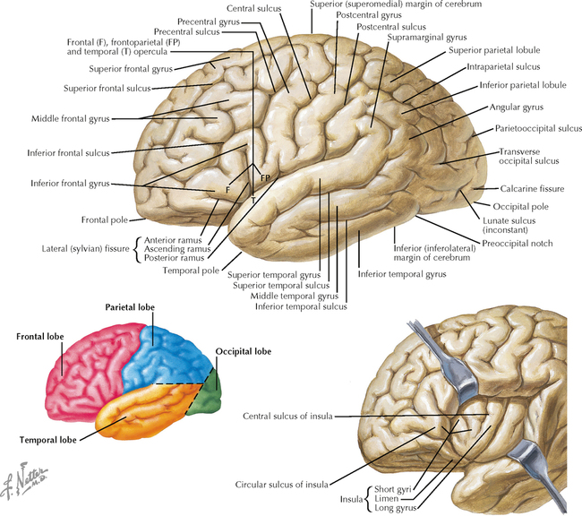
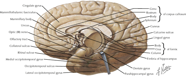
3 BRAIN 3.1. Surface Anatomy of the Forebrain: Lateral View 3.2. Lateral View of the Forebrain: Functional Regions 3.3. Lateral View of the Forebrain: Brodmann’s Areas 3.4. Anatomy of the Medial (Midsagittal) Surface of the Brain in Situ 3.5. Anatomy of the Medial (Midsagittal) Surface of the Brain, with Brain Stem Removed 3.6. Medial Surface of the Brain 3.7. Anatomy of the Basal Surface of the Brain, with the Brain Stem and Cerebellum Removed 3.8. Basal Surface of the Brain: Functional Areas and Brodmann’s Areas 3.9. Brain Imaging: Computed Tomography Scans, Coronal and Sagittal 3.10. Brain Imaging: Magnetic Resonance Imaging, Axial and Sagittal T1-Weighted Images 3.11. Brain Imaging: Magnetic Resonance Imaging, Axial and Sagittal T2-Weighted Images 3.12. Positron Emission Tomography Scanning 3.13. Horizontal Brain Sections Showing the Basal Ganglia 3.14. Major Limbic Forebrain Structures 3.15. Corpus Callosum 3.16. Color Imaging of the Corpus Callosum by Diffusion Tensor Imaging 3.17. Hippocampal Formation and Fornix 3.18. Thalamic Anatomy 3.19. Thalamic Nuclei 3.1 SURFACE ANATOMY OF THE FOREBRAIN: LATERAL VIEW The convolutions of the cerebral cortex allow a large expanse of cortex to be compactly folded into a small volume, an adaptation particularly prominent in primates. Major dependable landmarks separate the forebrain into lobes; the lateral (sylvian) fissure separates the temporal lobe below from the parietal and frontal lobes above, and the central sulcus separates the parietal and frontal lobes from each other. Several of the named gyri are associated with specific functional activities, such as the precentral gyrus (motor cortex) and the postcentral gyrus (primary sensory cortex). Some gyri, such as the superior, middle, and inferior frontal and temporal gyri, serve as anatomical landmarks of the cerebral cortex. The insula, the fifth lobe of the cerebral cortex, is deep to the outer cortex and can be seen by opening the lateral fissure. CLINICAL POINT Some functional characteristics of the cerebral cortex, such as long-term memory and some cognitive capabilities, cannot be localized easily to a particular gyrus or region of cortex. However, other functional capabilities are regionally localized. For example, the inferior frontal gyrus on the left contains the neuronal machinery for expressive language capabilities; the occipital pole, particularly along the upper and lower banks of the calcarine fissure, are specialized for visual processing from the retino-geniculo-calcarine system. Some very discrete lesions in further processing sites such as vision-related regions of the temporal lobe can result in specific deficits, such as agnosia for the recognition of faces or the inability to distinguish animate objects. This knowledge provides some clues about how feature extraction in sensory systems might be achieved in neuronal networks. 3.2 LATERAL VIEW OF THE FOREBRAIN: FUNCTIONAL REGIONS Some circumscribed regions of the cerebral hemisphere are associated with specific functional activities, including the motor cortex, the supplemental and premotor cortices, the frontal eye fields, the primary sensory cortex, and other association regions of the sensory cortex. Part of the auditory cortex is visible at the inferior edge of the lateral fissure (the transverse temporal gyrus of Heschl). Part of the visual cortex is visible at the occipital pole. Language areas of the left hemisphere include Broca’s area (expressive language) and Wernicke’s area (receptive language). Damage to these cortical regions results in loss of specific functional capabilities. There is some overlap between functional areas and named gyri (e.g., the motor cortex and the precentral gyrus), but there is no absolute concordance. CLINICAL POINT Some specific regions (gyri) of the cerebral cortex, such as the precentral gyrus (primary motor cortex) and the postcentral gyrus (primary somatosensory cortex), demonstrate topographic organization. Thus, information from the contralateral hand and arm are localized laterally, the body is represented more medially, and the lower extremity is represented along the midline and over the edge into the paracentral lobule. The face and head are represented in far lateral regions, just above the lateral fissure. This has important functional implications; damage to selected regions such as the midline territory, which is supplied with blood from the anterior cerebral artery, results in somatosensory loss and paresis in the contralateral lower extremity, while sparing the upper extremity. 3.3 LATERAL VIEW OF THE FOREBRAIN: BRODMANN’S AREAS Brodmann’s areas of the cerebral cortex have unique architectural characteristics in terms of the thickness and layering of the cerebral cortex; this knowledge is based on histological observations originally made by Korbinian Brodmann in 1909. His numbering of cortical areas is still used as a shorthand for describing the functional regions of the cortex, particularly those related to sensory functions. Some overlap exists among functional areas. For example, the motor cortex is area 4; the primary sensory cortex includes areas 3, 1, and 2; and the primary visual cortex is area 17. 3.4 ANATOMY OF THE MEDIAL (MIDSAGITTAL) SURFACE OF THE BRAIN IN SITU The entire extent of the neuraxis, from the spinomedullary junction through the brain stem, diencephalon, and telencephalon, is visible in a midsagittal section. The corpus callosum, a major commissural fiber bundle interconnecting the two hemispheres, is a landmark separating the cerebral cortex above from the thalamus, fornix, and subcortical forebrain below. The ventricular system, including the interventricular foramen (of Munro); the third ventricle (diencephalon); the cerebral aqueduct (midbrain); and the fourth ventricle (pons and medulla), is visible in a midsagittal view. This subarachnoid fluid system provides internal and external protection to the brain and also may serve as a fluid transport system for important regulatory molecules. The thalamus serves as a gateway to the cortex. The hypothalamic proximity to the median eminence (tuber cinereum) and the pituitary gland reflects the important role of the hypothalamus in regulating neuroendocrine function. A midsagittal view also reveals the midbrain colliculi, sometimes called the visual (superior) and auditory (inferior) tecta. CLINICAL POINT The right and left hemispheres are interconnected by commissural fiber bundles. The largest is the corpus callosum, which interconnects all lobes with their counterparts. The anterior commissure interconnects regions of the temporal lobes. When these commissural fiber bundles are disconnected (split brain), the hemispheres do not know what their counterparts are doing, and inputs to one hemisphere cannot produce an appropriate response from the opposite hemisphere. With a split brain, only a more generalized recognition of mood states occurs between the two hemispheres, presumably communicated through interconnections between lower structures, such as the diencephalon and brain stem. 3.5 ANATOMY OF THE MEDIAL (MIDSAGITTAL) SURFACE OF THE BRAIN, WITH BRAIN STEM REMOVED When the brain stem is removed, a midsagittal view reveals the C-shaped course of the fornix, extending from the hippocampal formation in the temporal lobe to the septum and hypothalamus. Temporal lobe structures, such as the parahippocampal cortex, the dentate gyrus and fimbria of the hippocampus, and the uncus (olfactory cortex) also are visible. In the hypothalamus, the caudal mammillary bodies and the interconnecting pathway to the thalamus, the mammillothalamic tract, are revealed. Only gold members can continue reading. Log In or Register to continue Share this:Click to share on Twitter (Opens in new window)Click to share on Facebook (Opens in new window) Related Related posts: VENTRICLES AND THE CEREBROSPINAL FLUID SPINAL CORD MOTOR SYSTEMS AUTONOMIC-HYPOTHALAMIC-LIMBIC SYSTEMS TELENCEPHALON PERIPHERAL NERVOUS SYSTEM Stay updated, free articles. Join our Telegram channel Join Tags: Netters Atlas of Neuroscience with STUDENT CONSULT Online Access Jun 4, 2016 | Posted by admin in NEUROLOGY | Comments Off on BRAIN Full access? Get Clinical Tree
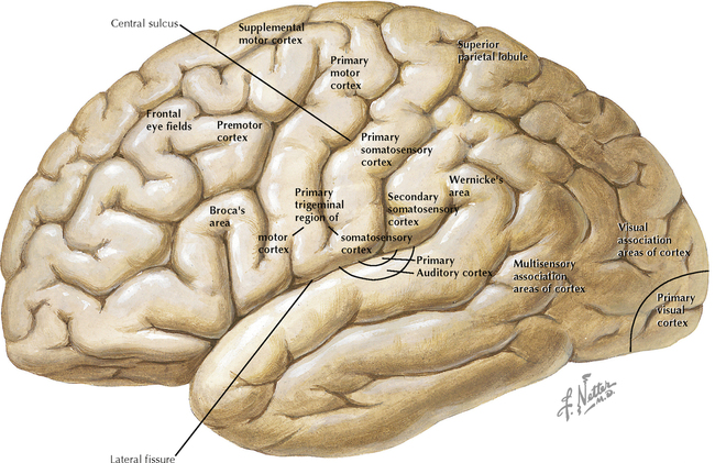
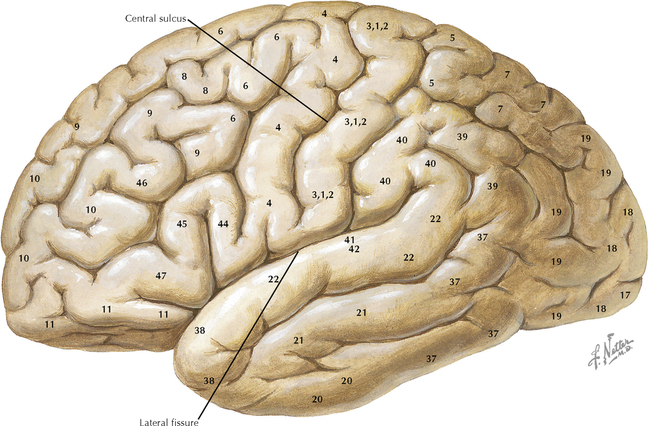
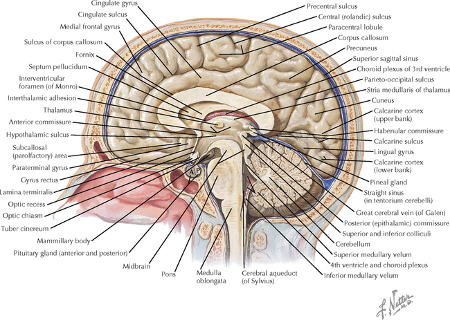
3 BRAIN 3.1. Surface Anatomy of the Forebrain: Lateral View 3.2. Lateral View of the Forebrain: Functional Regions 3.3. Lateral View of the Forebrain: Brodmann’s Areas 3.4. Anatomy of the Medial (Midsagittal) Surface of the Brain in Situ 3.5. Anatomy of the Medial (Midsagittal) Surface of the Brain, with Brain Stem Removed 3.6. Medial Surface of the Brain 3.7. Anatomy of the Basal Surface of the Brain, with the Brain Stem and Cerebellum Removed 3.8. Basal Surface of the Brain: Functional Areas and Brodmann’s Areas 3.9. Brain Imaging: Computed Tomography Scans, Coronal and Sagittal 3.10. Brain Imaging: Magnetic Resonance Imaging, Axial and Sagittal T1-Weighted Images 3.11. Brain Imaging: Magnetic Resonance Imaging, Axial and Sagittal T2-Weighted Images 3.12. Positron Emission Tomography Scanning 3.13. Horizontal Brain Sections Showing the Basal Ganglia 3.14. Major Limbic Forebrain Structures 3.15. Corpus Callosum 3.16. Color Imaging of the Corpus Callosum by Diffusion Tensor Imaging 3.17. Hippocampal Formation and Fornix 3.18. Thalamic Anatomy 3.19. Thalamic Nuclei 3.1 SURFACE ANATOMY OF THE FOREBRAIN: LATERAL VIEW The convolutions of the cerebral cortex allow a large expanse of cortex to be compactly folded into a small volume, an adaptation particularly prominent in primates. Major dependable landmarks separate the forebrain into lobes; the lateral (sylvian) fissure separates the temporal lobe below from the parietal and frontal lobes above, and the central sulcus separates the parietal and frontal lobes from each other. Several of the named gyri are associated with specific functional activities, such as the precentral gyrus (motor cortex) and the postcentral gyrus (primary sensory cortex). Some gyri, such as the superior, middle, and inferior frontal and temporal gyri, serve as anatomical landmarks of the cerebral cortex. The insula, the fifth lobe of the cerebral cortex, is deep to the outer cortex and can be seen by opening the lateral fissure. CLINICAL POINT Some functional characteristics of the cerebral cortex, such as long-term memory and some cognitive capabilities, cannot be localized easily to a particular gyrus or region of cortex. However, other functional capabilities are regionally localized. For example, the inferior frontal gyrus on the left contains the neuronal machinery for expressive language capabilities; the occipital pole, particularly along the upper and lower banks of the calcarine fissure, are specialized for visual processing from the retino-geniculo-calcarine system. Some very discrete lesions in further processing sites such as vision-related regions of the temporal lobe can result in specific deficits, such as agnosia for the recognition of faces or the inability to distinguish animate objects. This knowledge provides some clues about how feature extraction in sensory systems might be achieved in neuronal networks. 3.2 LATERAL VIEW OF THE FOREBRAIN: FUNCTIONAL REGIONS Some circumscribed regions of the cerebral hemisphere are associated with specific functional activities, including the motor cortex, the supplemental and premotor cortices, the frontal eye fields, the primary sensory cortex, and other association regions of the sensory cortex. Part of the auditory cortex is visible at the inferior edge of the lateral fissure (the transverse temporal gyrus of Heschl). Part of the visual cortex is visible at the occipital pole. Language areas of the left hemisphere include Broca’s area (expressive language) and Wernicke’s area (receptive language). Damage to these cortical regions results in loss of specific functional capabilities. There is some overlap between functional areas and named gyri (e.g., the motor cortex and the precentral gyrus), but there is no absolute concordance. CLINICAL POINT Some specific regions (gyri) of the cerebral cortex, such as the precentral gyrus (primary motor cortex) and the postcentral gyrus (primary somatosensory cortex), demonstrate topographic organization. Thus, information from the contralateral hand and arm are localized laterally, the body is represented more medially, and the lower extremity is represented along the midline and over the edge into the paracentral lobule. The face and head are represented in far lateral regions, just above the lateral fissure. This has important functional implications; damage to selected regions such as the midline territory, which is supplied with blood from the anterior cerebral artery, results in somatosensory loss and paresis in the contralateral lower extremity, while sparing the upper extremity. 3.3 LATERAL VIEW OF THE FOREBRAIN: BRODMANN’S AREAS Brodmann’s areas of the cerebral cortex have unique architectural characteristics in terms of the thickness and layering of the cerebral cortex; this knowledge is based on histological observations originally made by Korbinian Brodmann in 1909. His numbering of cortical areas is still used as a shorthand for describing the functional regions of the cortex, particularly those related to sensory functions. Some overlap exists among functional areas. For example, the motor cortex is area 4; the primary sensory cortex includes areas 3, 1, and 2; and the primary visual cortex is area 17. 3.4 ANATOMY OF THE MEDIAL (MIDSAGITTAL) SURFACE OF THE BRAIN IN SITU The entire extent of the neuraxis, from the spinomedullary junction through the brain stem, diencephalon, and telencephalon, is visible in a midsagittal section. The corpus callosum, a major commissural fiber bundle interconnecting the two hemispheres, is a landmark separating the cerebral cortex above from the thalamus, fornix, and subcortical forebrain below. The ventricular system, including the interventricular foramen (of Munro); the third ventricle (diencephalon); the cerebral aqueduct (midbrain); and the fourth ventricle (pons and medulla), is visible in a midsagittal view. This subarachnoid fluid system provides internal and external protection to the brain and also may serve as a fluid transport system for important regulatory molecules. The thalamus serves as a gateway to the cortex. The hypothalamic proximity to the median eminence (tuber cinereum) and the pituitary gland reflects the important role of the hypothalamus in regulating neuroendocrine function. A midsagittal view also reveals the midbrain colliculi, sometimes called the visual (superior) and auditory (inferior) tecta. CLINICAL POINT The right and left hemispheres are interconnected by commissural fiber bundles. The largest is the corpus callosum, which interconnects all lobes with their counterparts. The anterior commissure interconnects regions of the temporal lobes. When these commissural fiber bundles are disconnected (split brain), the hemispheres do not know what their counterparts are doing, and inputs to one hemisphere cannot produce an appropriate response from the opposite hemisphere. With a split brain, only a more generalized recognition of mood states occurs between the two hemispheres, presumably communicated through interconnections between lower structures, such as the diencephalon and brain stem. 3.5 ANATOMY OF THE MEDIAL (MIDSAGITTAL) SURFACE OF THE BRAIN, WITH BRAIN STEM REMOVED When the brain stem is removed, a midsagittal view reveals the C-shaped course of the fornix, extending from the hippocampal formation in the temporal lobe to the septum and hypothalamus. Temporal lobe structures, such as the parahippocampal cortex, the dentate gyrus and fimbria of the hippocampus, and the uncus (olfactory cortex) also are visible. In the hypothalamus, the caudal mammillary bodies and the interconnecting pathway to the thalamus, the mammillothalamic tract, are revealed. Only gold members can continue reading. Log In or Register to continue Share this:Click to share on Twitter (Opens in new window)Click to share on Facebook (Opens in new window) Related Related posts: VENTRICLES AND THE CEREBROSPINAL FLUID SPINAL CORD MOTOR SYSTEMS AUTONOMIC-HYPOTHALAMIC-LIMBIC SYSTEMS TELENCEPHALON PERIPHERAL NERVOUS SYSTEM Stay updated, free articles. Join our Telegram channel Join Tags: Netters Atlas of Neuroscience with STUDENT CONSULT Online Access Jun 4, 2016 | Posted by admin in NEUROLOGY | Comments Off on BRAIN Full access? Get Clinical Tree