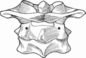
13 Kuniyoshi Abumi To place a screw safely into the cervical pedicle with avoidance of injury to the surrounding neurovascular structures. Cervical pedicle screws have nearly twice the pullout strength of lateral mass screws. In addition, cervical pedicle screw instrumentation provides a greater stabilization effect than other cervical internal fixation procedures. Cervical pedicle screws may be used in several settings in which the cervical lamina or lateral masses are inadequate because of fracture, tumor destruction, postsurgical disturbances, or marked osteoporosis. Cervical pedicle screw fixation, due to its rigid anchorage within the cervical pedicle, provides an opportunity for greater correctability of kyphotic deformity than lateral mass screw fixation. Preoperative imaging studies include computed tomography (CT) to help delineate bony anatomy. Reconstructive CT in the oblique plane provides useful information on the size of the neural foramen. Magnetic resonance imaging (MRI) and magnetic resonance angiography facilitate determining the precise location of the vertebral arteries. In about 5% of the population, the foramen transversarium of C7 contains the vertebral artery. This should be appreciated prior to pedicle screw insertion. The patient is placed in the prone position with a Mayfield headrest and shoulder taping to pull the shoulder girdles caudally for proper lateral fluoroscopy image. Intraoperative lateral fluoroscopy is helpful to determine the level of screw entry point in the sagittal plane, screw depth, and trajectory. Laminoforaminotomy of the subaxial vertebrae allows tactile identification of the cephalad, medial, and caudad pedicle borders, especially for the C7 pedicle when adequate lateral fluoroscopic image may not be available. An alternative fixation method or possibly unilateral pedicle screw fixation should be considered if a pedicle breach is encountered. The starting point of C2 pedicle screw placement is the cranial medial portion of the C2 lateral mass (Fig. 13.1). A nerve retractor should be placed within the canal to confirm the medial border of the C2 isthmus and pedicle. A 2- to 3-mm high-speed diamond burr may be used to create an entrance hole on the lateral mass of C2. A pedicle probe insertion is recommended prior to tapping and screwing. The direction is approximately 15 to 20 degrees medially in the horizontal plane (Fig. 13.2) and perpendicular to the anterior surface of the axis (Fig. 13.3). The ventral cortex of C2 may be penetrated by the screw.
Cervical Pedicle Screw Placement
Description
Key Principles
Expectations
Indications
Contraindications
Special Considerations
Special Instructions, Position, and Anesthesia
Tips, Pearls, and Lessons Learned
Difficulties Encountered
Key Procedural Steps
Placement of C2
Related posts:


Stay updated, free articles. Join our Telegram channel

Full access? Get Clinical Tree


