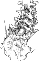
52 Michael E. Janssen and Jason C. Datta Placement of an interbody fibular strut graft through the sacrum and L5 vertebral body for high-grade lumbosacral spondylolisthesis. Placement of the strut graft fulfills the following goals in the surgical treatment of high-grade spondylolisthe-sis: immediate structural support and stability, solid bony interface for fusion, and a graft placed under compression. The posterior placement of the graft requires a large decompression of the neural elements, which is advantageous in patients with neurologic complaints. Spinal fusion usually occurs within 3 to 6 months. High-grade lumbosacral spondylolisthesis greater than Meyerding grade III that has failed nonoperative treatment for mechanical back pain, lumbosacral neurologic dysfunction, slip progression, or revision of previous failed surgery. This procedure may be ideal for revision surgeries where the posterolateral graft bed is poor, or in the setting of a previously anterior bony strut graft procedure. The posterior procedure is limited by the cephalad orientation of the graft tract by the patient’s body. This limits the procedure to an in-situ fusion of a slip of greater than 50% or the partial reduction of a higher grade slip to 50%. Presurgical radiographic evaluation for preoperative planning for both these procedures is essential. This includes preoperative plain films in the upright position to evaluate the posterior element dysplasia and slip angle. This defines lamina and pedicle dysplasia identifying areas at risk during decompression, and the planning of safe segmental posterior stabilization. Dysplasia of the transverse processes of L4 and L5 should be evaluated for the surface area available for posterolateral fusion. Transverse process surface area <2 cm 2 has been correlated with a high pseudarthrosis rate. It is strongly recommended to add the stability and superior graft bed of an interbody strut graft at the lumbosacral junction. Magnetic resonance imaging (MRI) or computed tomography (CT) myelography should be used to evaluate the geometry and pathology of neural compression. The presence of sacral inlet stenosis should alert the surgeon to the need of a sacral dome resection. Regardless of sacral inlet stenosis, sacral dome resection may ease reduction forces required and the strain placed on the L5 nerve roots by columnar shortening. CT angiography may be a useful adjunct to define the prevertebral vascular anatomy prior to an anterior procedure in a high-grade deformity. The patient should be placed in the prone position on a radiolucent four-poster frame. Intraoperative fluoroscopy is required. The graft used, autograft versus allograft, does not appear to affect outcomes in the limited literature available. There may be a role for a vascularized fibular graft in previously radiated tissues. Reduction forces may be decreased by a wide thorough decompression, sacroplasty, and slight distraction. Resection of the iliolumbar ligament and far lateral foraminal decompression may decrease the rate of neurologic complications from the L5 root during reduction maneuvers. A cannulated reamer from an anterior cruciate ligament reconstruction set is ideal for both the anterior and posterior procedures. An appropriate-size reamer can be chosen using the graft sizing tubes over the strut graft. The reamer sleeves in these sets serve as an excellent soft tissue protector. Both the procedures can be used without supplemental posterior segmental instrumentation, but it is felt that higher rates of successful fusion can be achieved with instrumentation. The added instrumentation is needed with dysplastic transverse processes <2 cm 2. This includes L4 pedicle screws, bilateral bicorti-cal S1 fixation, and iliac screws measuring >60 mm in length and 7 mm in diameter to improve fusion rates and decrease interbody strut graft failure. The posterior placement of a strut graft requires a standard midline approach to the lumbosacral spine with exposure of the posterolateral gutters from the L4 transverse process to the sacral ala. Decompression includes partial laminectomy of the inferior portion of L4 and complete laminectomy of L5, S1, and S2 (Fig. 52.1). Wide foraminal decompression of the L5 nerve roots is required. Sacroplasty is needed in the setting of sacral inlet stenosis, and may be performed to ease reduction. This is best achieved with a broad curved osteotome under direct visualization. If instrumentation is to be used, it should be placed prior to reduction or graft placement. This typically requires fixation from L4 to the sacrum and in most instances the pelvis.
Lumbosacral Interbody Fibular Strut Placement for High-Grade Spondylolisthesis: Anterior Speed’s Procedure and Posterior Procedure
Description
Key Principles
Indications
Contraindications
Special Considerations
Special Instructions, Position, and Anesthesia
Tips, Pearls, and Lessons Learned
Key Procedural Steps
Related posts:
 Anterior Thoracic and Thoracolumbar Plating Techniques
 Anterior Cervical Corpectomy
 Posterior Spinal Anchor Strategy Placement and Rod Reduction Techniques: Posterior (Rotation vs. In-Situ Translation)
 Iliosacral Screw Fixation Techniques
 Open Lumbar Microscopic Diskectomy
 Spondylolysis Repair (Pars Interarticularis Repair)
Stay updated, free articles. Join our Telegram channel

Full access? Get Clinical Tree


