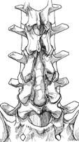
36 Mark A. Knaub and Jeffrey S. Fischgrund To safely perform decompression of the neural elements in the lumbar spine below the level of the conus medullaris without damage to the neurologic structures. Decompression of the spinal canal and foramen via laminectomy, partial facetectomy, and foraminotomy can be safely performed without injury to the neurologic structures and dural sac while maintaining the stability of the lumbar motion segments involved. Decompressive laminectomy and facetectomy for spinal stenosis relieves symptoms of neurogenic claudication and radiculopathy in a vast majority of patients. Carefully performed surgery leads to few complications, a short hospital stay, and a rapid improvement in the patient’s symptoms. Patients can expect to return to their activities of daily living in a relatively short period of time. Patients with neurogenic claudication or radiculopathy with confirmatory imaging studies documenting spinal stenosis. Preoperative imaging studies must show evidence of neurologic compression that corresponds to the patient’s symptoms and signs. A complete understanding of the local anatomy contributing to the patient’s stenosis is mandatory to thoroughly decompress the neural elements. Elderly patients should have routine medical evaluation and optimization to lessen the likelihood of perioperative medical complications. Regional anesthesia via a spinal or general anesthesia can be utilized. The patient is placed in the prone position with the hips and knees flexed. Alternatively, the patient may be placed in a kneeling position. Flexion of the hips allows for straightening of the lumbar lordosis and opening of the interlaminar window, which will make decompression easier. For each of these positions the abdomen should hang free, which decreases the intraabdominal pressure and therefore the central venous pressure, leading to decreased engorgement of the epidural venous plexus. This decreases intraoperative blood loss. Careful positioning of the head and extremities should be employed to avoid iatrogenic neurologic injury or pressure-related problems. Special attention should be given to the eyes/orbits, especially when using general anesthesia. If spinal anesthesia is employed the puncture must be cranial to the level of planned decompression if possible. A cerebrospinal fluid leak may develop postoperatively if the puncture site is uncovered by the decompression. Intraoperative imaging is mandatory to confirm the correct level of decompression. The use of anatomic landmarks can be helpful in many cases, but normal variants, such as sacralization of the lowest lumbar segment, may lead to confusion and ultimately to wrong-level surgery. Visualization can be enhanced with an operating microscope or with a combination of loupes and a fiber-optic head light. If limited imaging studies are available, special care must be taken to avoid inadequate decompression. Computed tomography (CT) scans frequently only include from L3 to S1, so additional imaging may be necessary to ensure that the upper lumbar segments are not involved in the pathology. A double puncture technique may be necessary in patients with complete myelographic blocks to adequately assess the segments cranial to the block. Patients with severe stenosis or synovial cysts often have significant adhesions between the ligamentum flavum and the dural sac. Elderly patients tend to have a thinner, more fragile dural sac. Meticulous surgical technique should be employed to avoid an incidental durotomy. Once the levels to be included in the decompression have been identified with intraoperative imaging, the interspinous ligament of the involved segments is removed with a large Leksell rongeur. This rongeur is then used to remove half of the inferior spinous process and half of the most superior spinous process. After thinning the lamina with a rongeur or a high-speed burr, the central portion of the canal is decompressed (Fig. 36.1). Decompression is generally carried out from a caudal to cranial direction with Kerrison rongeurs. The ligamentum flavum may be temporarily left in place to protect the underlying dura. The location of the lateral edge of the pars interarticularis must be known to avoid iatrogenic pars removal or thinning of the pars, which may predispose the patient to a fracture.
Open Laminectomy, Medial Facetectomy, and Foraminotomy
Description
Key Principles
Expectations
Indications
Contraindications
Special Considerations
Special Instructions, Position, and Anesthesia
Tips, Pearls, and Lessons Learned
Difficulties Encountered
Key Procedural Steps
Related posts:


Stay updated, free articles. Join our Telegram channel

Full access? Get Clinical Tree


