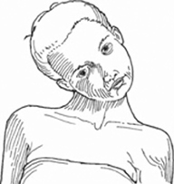
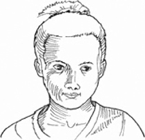
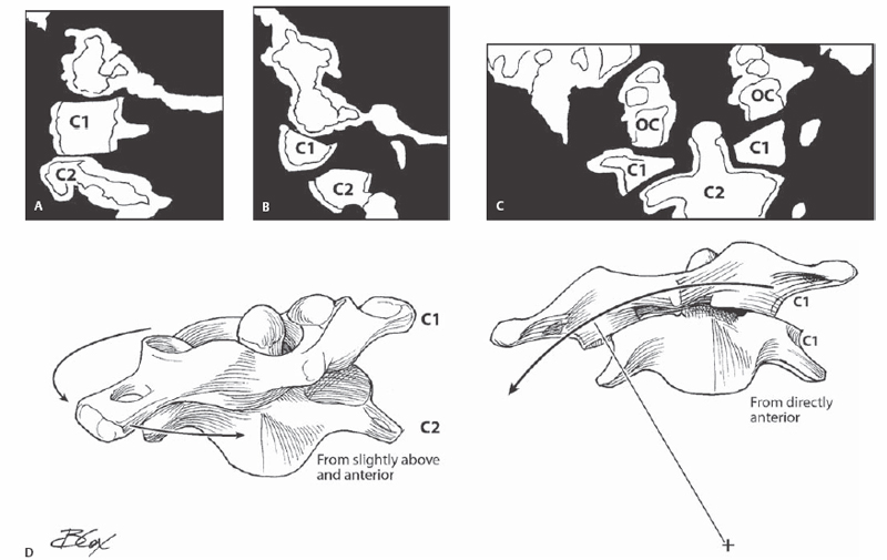
9 Rick C. Sasso Atlantoaxial rotatory subluxation is an anterior displacement of one C1-C2 joint with a concomitant posterior migration of the contralateral articulation. The pivot point is usually an intact anterior arch of C1-odontoid articulation with a preserved transverse atlantal ligament (TAL). This type of C1-C2 subluxation is different from the more common anterior subluxation of C1-C2, which routinely occurs due to incompetence of the TAL. Rheumatoid involvement and trauma to the TAL are the most common reasons for anterior subluxation of the atlantoaxial joint. In a different type of atlantoaxial rotatory subluxation, the pivot point is one C1-C2 joint, whereas the contralateral C1 lateral mass is shifted anterior to C2. The TAL is usually attenuated or frankly disrupted in this variety of rotatory subluxation. Atlantoaxial rotatory subluxation usually occurs in children and is a common cause of torticollis (Fig. 9.1). This is routinely caused by trauma or infection of the upper respiratory tract, which weakens the ligamentous and facet capsular structures of C1-C2. The flat C1-C2 facet joints do not provide intrinsic stability and are specialized to allow rotation. Minimal trauma and minor pharyngeal inflammation can therefore lead to C1–C2 rotatory subluxation. Fig. 9.1 A 10-year-old girl 5 months after a car crash with a fixed torticollis. This severe deformity continued despite multiple attempts of closed reduction and immobilization even with a halo. Most C1-C2 rotatory subluxations in children reduce spontaneously. The torticollis deformity may be treated with a short course of a cervical collar or low-weight traction with a head halter. If these maneuvers do not reduce the deformity, then open reduction and internal fixation is contemplated. The articulation is best visualized, reduced, instrumented, and fused from a posterior approach. The most powerful method of reduction and instrumentation is through anchoring a screw into each lateral mass of C1 and pedicles of C2 to directly manipulate the vertebrae with these “joysticks.” Anatomic reduction of the atlantoaxial articulation with resolution of the torticollis deformity is expected (Fig. 9.2). With direct access to the joints and focused manipulation through the segmental fixation into C1 and C2, perfect reduction is anticipated. Fig. 9.2 The same girl as in Fig. 9.1 one day after open reduction and internal fixation of the C1-C2 joints. The indications for this technique of direct reduction and internal fixation with polyaxial lateral mass screws in C1 and pedicles screws in C2 connected through rods are irreducible rotatory subluxations of C1-C2 that are unresponsive to nonoperative treatment (Fig. 9.3).
Reduction Techniques for Atlantoaxial Rotary Subluxation
Description
Expectations
Indications
Related posts:
 Anterior Thoracic and Thoracolumbar Plating Techniques
 Anterior Cervical Corpectomy
 Posterior Spinal Anchor Strategy Placement and Rod Reduction Techniques: Posterior (Rotation vs. In-Situ Translation)
 Iliosacral Screw Fixation Techniques
 Open Lumbar Microscopic Diskectomy
 Spondylolysis Repair (Pars Interarticularis Repair)
Stay updated, free articles. Join our Telegram channel

Full access? Get Clinical Tree


