Chapter 5 Stupor and Coma
Definitions
Consciousness may be defined as a state of awareness of self and surroundings. Alterations in consciousness are conceptualized into two types. The first type affects arousal and is the subject of this chapter. The second type involves cognitive and affective mental function, sometimes referred to as the “content” of mental function. Examples of the latter type of alteration in consciousness are dementia (see Chapter 6), delusions, confusion, and inattention (see Chapter 9). These altered states of consciousness, with the exception of advanced dementia, do not affect the level of arousal. Sleep, the only normal form of altered consciousness, is discussed in Chapter 68.
Conditions That May Mimic Coma
Several different states of impaired cognition or consciousness may appear similar to coma or be confused with it (Table 5.1). Moreover, patients who survive the initial coma may progress to certain of these syndromes after varying lengths of time. Once sleep/wake cycles become established, true coma is no longer present. Differentiation of these states from true coma is important to allow administration of appropriate therapy and help determine prognosis.
In the locked-in syndrome (de-efferented state), patients are alert and aware of their environment but are quadriplegic, with lower cranial nerve palsies resulting from bilateral ventral pontine lesions that involve the corticospinal, corticopontine, and corticobulbar tracts. These patients are awake and alert but are voluntarily able only to move their eyes vertically or blink. The locked-in syndrome most often is observed as a consequence of pontine infarction due to basilar artery thrombosis. Other causes include central pontine myelinolysis and brainstem mass lesions. A state similar to the locked-in syndrome also may be seen with severe polyneuropathy—in particular, acute inflammatory demyelinating polyradiculoneuropathy, myasthenia gravis, and poisoning with neuromuscular blocking agents.
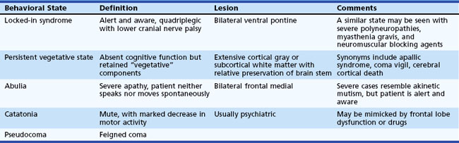
In the persistent vegetative state (PVS), patients have lost cognitive neurological function but retain vegetative or noncognitive neurological function such as cardiac action, respiration, and maintenance of blood pressure. This state follows coma and is characterized by absence of cognitive function or awareness of the environment despite a preserved sleep/wake cycle. Spontaneous movements may occur, and the eyes may open in response to external stimuli, but the patient does not speak or obey commands. Diagnostic criteria for PVS are provided in Box 5.1. Diagnosis of this condition should be made cautiously and only after extended periods of observation. A number of poorly defined syndromes have been used synonymously with PVS, including apallic syndrome or state, akinetic mutism, coma vigil, alpha coma, neocortical death, and permanent unconsciousness. These terms, used variously by different authors, probably are best avoided because of their lack of precision.
Box 5.1
Criteria for Diagnosis of Persistent Vegetative State
1. No evidence of awareness of themselves or their environment; they are incapable of interacting with others
2. No evidence of sustained, reproducible, purposeful, or voluntary behavioral responses to visual, auditory, tactile, or noxious stimuli
3. No evidence of language comprehension or expression
4. Intermittent wakefulness manifested by the presence of sleep/wake cycles
5. Sufficiently preserved hypothalamic and brainstem autonomic functions to survive if given medical and nursing care
6. Bowel and bladder incontinence
7. Variably preserved cranial nerve (pupillary, oculocephalic, corneal, vestibulo-ocular, and gag) and spinal reflexes
Data from The Multi-Society Task Force on PVS, 1994. Medical aspects of the persistent vegetative state. N Engl J Med 330, 1499-1508, 1572-1579.
A condition that has been estimated to be 10 times more common than PVS is the minimally conscious state, in which severe disability accompanies minimal awareness. A set of diagnostic criteria for the minimally conscious state has been proposed (Box 5.2). Abulia is a severe apathy in which patients have blunting of feeling, drive, mentation, and behavior such that they neither speak nor move spontaneously.
Box 5.2
Criteria for the Minimally Conscious State
To diagnose a minimally conscious state, limited but clearly discernible evidence of self- or environmental awareness must be demonstrated on a reproducible or sustained basis by one or more of the following behaviors:
2. Gestural or verbal yes/no responses (regardless of accuracy)
4. Purposeful behavior, including movements or affective behaviors that occur in contingent relationship to relevant environmental stimuli and are not due to reflexive activity
Data from Giacino, J.T., Ashwal, S., Childs, N., et al., 2002. The minimally conscious state: definition and diagnostic criteria. Neurology 58, 349-353.
Catatonia may result in a state of muteness with dramatically decreased motor activity. The maintenance of body posture, with preserved ability to sit or stand, distinguishes it from organic pathological stupor. It generally is a psychiatric manifestation but may be mimicked by frontal lobe dysfunction or drug effect.
Pseudocoma is the term for a condition in which the patient appears comatose (i.e., unresponsive, unarousable, or both) but has no structural, metabolic, or toxic disorder.
Approach to the Patient in Coma
The initial clinical approach to the patient in a state of stupor or coma is based on the principle that all alterations in arousal constitute acute life-threatening emergencies until vital functions such as blood pressure and oxygenation are stabilized, potentially reversible causes of coma are treated, and the underlying cause of the alteration in arousal is understood. Urgent steps may be necessary to avoid or minimize permanent brain damage from reversible causes. In view of the urgency of this situation, every physician should develop a diagnostic and therapeutic routine to use with a patient with an alteration in consciousness. A basic understanding of the mechanisms that lead to impairment in arousal is necessary to develop this routine. The anatomical and physiological bases for alterations in arousal are discussed in Chapter 68.
Although it is essential to keep in mind the concept of a spectrum of arousal, for the sake of simplicity and brevity only the term coma is used in the rest of this chapter. Table 5.2 lists many of the common causes of coma. More than half of all cases of coma are due to diffuse and metabolic brain dysfunction. In Plum and Posner’s landmark study (1980, see 2007 revision) of 500 patients initially diagnosed as having coma of unknown cause (in whom the diagnosis was ultimately established), 326 patients had diffuse and metabolic brain dysfunction. Almost half of these had drug poisonings. Of the remaining patients, 101 had supratentorial mass lesions, including 77 hemorrhagic lesions and 9 infarctions; 65 had subtentorial lesions, mainly brainstem infarctions; and 8 had psychiatric coma.
I. SYMMETRICAL-NONSTRUCTURAL |
Toxins |
Drugs |
II. SYMMETRICAL-STRUCTURAL |
Supratentorial |
III. ASYMMETRICAL-STRUCTURAL |
Supratentorial |
Metabolic |
Subarachnoid Hemorrhage |
Subdural Hemorrhage, Bilateral |
Infections |
Psychiatric |
Catatonia |
Other |
Infratentorial |
Subdural Empyema |
* Relatively common asymmetrical presentation.
Data from Plum, F., Posner, J.B., 1980. The Diagnosis of Stupor and Coma, third ed. F.A. Davis, Philadelphia; and from Fisher, C.M., 1969. The neurological examination of the comatose patient. Acta Neurol Scand 45, 1-56.
A logical decision tree often used in searching for the cause of coma divides the categories of diseases that cause coma into three groups: structural lesions, which may be above or below the tentorium; metabolic and toxic causes; and psychiatric causes. The history and physical examination usually provide sufficient evidence to determine the presence or absence of a structural lesion and quickly differentiate the general categories to either decide what further diagnostic tests are needed or allow for immediate intervention if necessary.
Serial examinations are needed, with precise description of the behavioral state at different points in time to determine whether the patient is improving or—a more ominous finding—worsening, and to decide whether a change in therapy or further diagnostic testing is necessary. Subtle declines in the intermediate states of arousal may herald precipitous changes in brainstem function, which may affect regulation of vital functions such as respiration or blood pressure. The dynamic quality of alterations of consciousness and the need for accurate documentation at different points in time cannot be overemphasized.
Rapid Initial Examination and Emergency Therapy
A relatively quick initial assessment is conducted to ensure that the comatose patient is medically and neurologically stable before a more detailed investigation is undertaken. This rapid initial examination is essential to rule out the need for immediate medical or surgical intervention. In addition, various supportive or preventive measures may be indicated.
Urgent and sometimes empirical therapy is given to prevent further brain damage. Potential immediate metabolic needs of the brain are supplied by empirical use of supplemental oxygen, intravenous (IV) thiamine (at least 100 mg), and IV 50% dextrose in water (25 g). A baseline serum glucose level should be obtained before glucose administration.
The use of IV glucose in patients with ischemic or anoxic brain damage is controversial. Extra glucose may augment local lactic acid production by anaerobic glycolysis and may worsen ischemic or anoxic damage. Clinically, however, we currently recommend empirical glucose administration when the cause of coma is unknown. There are two reasons for this approach: (1) the frequent occurrence of alterations in arousal due to hypoglycemia and the relatively good prognosis for coma due to hypoglycemia when it is treated expeditiously; and (2) the potentially permanent consequences if it is not treated. By comparison, the prognosis for anoxic or ischemic coma generally is poor and probably will remain poor regardless of glucose supplementation.
Thiamine must always be given in conjunction with glucose to prevent precipitation of Wernicke encephalopathy. Naloxone hydrochloride may be given parenterally, preferably IV, in doses of 0.4 to 2 mg if opiate overdose is the suspected cause of coma. An abrupt and complete reversal of narcotic effect may precipitate an acute abstinence syndrome in persons who are physically dependent on opiates.
Initial examination should include a check of general appearance, blood pressure, pulse, temperature, respiratory rate and breath sounds, best response to stimulation, pupil size and responsiveness, and posturing or adventitious movements. The neck should be stabilized in all instances of trauma until cervical spine fracture or subluxation can be ruled out. The airway should be protected in all comatose patients and an IV line placed.
In coma, however, the classic sign of an acute condition in the abdomen—namely, abdominal rigidity—may be subtle or absent. In addition, the diagnosis of blunt abdominal trauma is difficult in patients with a change in mental status. Therefore, in unconscious patients with a history of trauma, peritoneal lavage by an experienced surgeon may be warranted.
Hypotension, marked hypertension, bradycardia, arrhythmias causing depression of blood pressure, marked hyperthermia, and signs of cerebral herniation mandate immediate therapeutic intervention.
Hyperthermia or meningismus prompts consideration of urgent lumbar puncture (LP). Examination of the fundus of the eye for papilledema and a computed tomography (CT) scan of the brain should be performed before LP in any comatose patient. Although the only absolute contraindication to LP is the presence of an infection over the site of puncture, medicolegal considerations make a CT scan mandatory before LP. To avoid a delay in therapy required to perform a CT scan, some authorities recommend initiating antibiotics immediately when acute bacterial meningitis is strongly suspected, though this may prevent subsequent identification of the responsible organism.
The risk of herniation from an LP in patients with evidence of increased intracerebral pressure is difficult to ascertain from the literature; estimates range from 1% to 12%, depending on the series (Posner et al., 2007). It is important to recognize that both central and tonsillar herniation may increase neck tone.
Ecchymosis, petechiae, or evidence of ready bleeding on general examination may indicate coagulation abnormality or thrombocytopenia. This increases the risk of epidural hematoma after an LP, which may cause devastating spinal cord compression. Measurements of prothrombin time, partial thromboplastin time, and platelet count should precede LP in these cases, and the coagulation abnormality or thrombocytopenia should be corrected before proceeding to LP.
Common Presentations
Coma usually manifests in one of three ways. Most commonly, it occurs as an expected or predictable progression of an underlying illness. Examples are focal brainstem infarction with extension; chronic obstructive pulmonary disease in a patient who is given too high a concentration of oxygen, thereby decreasing respiratory drive and resulting in carbon dioxide narcosis; and known barbiturate overdose when the ingested drug cannot be fully removed and begins to cause unresponsiveness. Second, coma occurs as an unpredictable event in a patient whose prior medical conditions are known to the physician. The coma may be a complication of an underlying medical illness, such as in a patient with arrhythmia who suffers anoxia after a cardiac arrest. Alternatively, an unrelated event may occur, such as sepsis from an IV line in a cardiac patient or stroke in a hypothyroid patient. Finally, coma can occur in a patient whose medical history is totally unknown to the physician. Sometimes this type of presentation is associated with a known probable cause such as head trauma incurred in a motor vehicle accident, but often the unknown comatose patient presents to the physician without an obvious associated cause. Although the patient without an obvious cause of coma may seem most challenging, thorough objective systematic assessment must be applied in every comatose patient. Special care must be taken not to be lulled or misled by an apparently predictable progression of an underlying illness or other obvious cause of coma.
History
Once the patient is relatively stable, clues to the cause of the coma should be sought by briefly interviewing relatives, friends, bystanders, or medical personnel who may have observed the patient before or during the decrease in consciousness. Telephone calls to family members may be helpful. The patient’s wallet or purse should be examined for lists of medications, a physician’s card, or other information.
Attempts should be made to ascertain the patient’s social background and prior medical history and the circumstances in which the patient was found. The presence of drug paraphernalia or empty medicine bottles suggests a drug overdose. Newer recreational drugs, such as γ-hydroxybutyrate (GHB), must be considered in the differential diagnosis. An oral hypoglycemic agent or insulin in the medicine cabinet or refrigerator implies possible hypoglycemia. Antiarrhythmic agents such as procainamide or quinidine suggest existing coronary artery disease with possible myocardial infarction (MI) or warn that an unwitnessed arrhythmia may have caused cerebral hypoperfusion, with resulting anoxic encephalopathy. Warfarin, typically prescribed for patients with deep venous thrombosis or pulmonary embolism, those at risk for cerebral embolism, and those with a history of brainstem or cerebral ischemia, may be responsible for massive intracerebral bleeding. In patients found to be unresponsive at the scene of an accident such as a car crash, the unresponsive state may be due to trauma incurred in the accident, or sudden loss of consciousness may have precipitated the accident.
The neurologist often is called when patients do not awaken after surgery or when coma supervenes following a surgical procedure. Postoperative causes of coma include many of those listed in Table 5.4. In addition, the physician also must have a high index of suspicion for certain neurological conditions that occur in this setting, including fat embolism, addisonian crisis, hypothyroid coma (precipitated by acute illness or surgical stress), Wernicke encephalopathy from carbohydrate loading without adequate thiamine stores, and iatrogenic overdose of a narcotic analgesic.
Finally, family members or friends may be invaluable in identifying psychiatric causes of unresponsiveness. The family may describe a long history of psychiatric disease, previous similar episodes from which the patient recovered, current social stresses on the patient, or the patient’s unusual idiosyncratic response to stress. Special care must be taken with psychiatric patients because of the often biased approach to these patients, which may lead to incomplete evaluation. Psychiatric patients are subject to all the causes of coma listed in Table 5.4.
General Examination
A systematic, detailed general examination is especially helpful in the approach to the comatose patient who is unable to describe prior or current medical problems. This examination begins in the initial rapid examination with evaluation of blood pressure, pulse, respiratory rate, and temperature.
Blood Pressure Evaluation
Hypotension
Cerebral hypoperfusion secondary to hypotension may result in coma if the mean arterial pressure falls below the value for which the brain is able to autoregulate (normally 60 mm Hg). This value is substantially higher in chronically hypertensive persons, in whom the cerebral blood flow–mean arterial pressure curve is shifted to the right. Among the causes of hypotension are hypovolemia, massive external or internal hemorrhage, MI, cardiac tamponade, dissecting aortic aneurysm, intoxication with alcohol or other drugs (especially barbiturates), toxins, Wernicke encephalopathy, Addison disease, and sepsis. Although most patients with hypotension are cold because of peripheral vasoconstriction, patients with Addison disease or sepsis may have warm shock due to peripheral vasodilation. Medullary damage also may result in hypotension due to damage to the pressor center.
Hypertension
Hypertension is the cause of alterations in arousal in hypertensive crisis and is seen secondarily as a response to cerebral infarction, in subarachnoid hemorrhage, with certain brainstem infarctions, and with increased intracerebral pressure. The Kocher-Cushing (or Claude Bernard) reflex is the development of hypertension associated with bradycardia and respiratory irregularity due to increased ICP. This response occurs more commonly in the setting of a posterior fossa lesion and in children. It results from compression or ischemia of the pressor area lying beneath the floor of the fourth ventricle. Hypertension is a common condition and thus may be present but unrelated to the cause of coma.
Heart Rate
In addition to the Kocher-Cushing reflex, bradycardia can result from myocardial conduction blocks, with certain poisonings, and from effects of drugs such as the beta-blockers. Tachycardia is a result of hypovolemia, hyperthyroidism, fever, anemia, and certain toxins and drugs including cocaine, atropine, and other anticholinergic medications.
Respiration
The most common causes of decreased respiratory rate are metabolic or toxic, such as carbon dioxide narcosis or drug overdose with central nervous system (CNS) depressants. Increased respiratory rate can result from hypoxia, hypercapnia, acidosis, hyperthermia, hepatic disease, toxins or drugs (especially those that produce metabolic acidosis, such as methanol, ethylene glycol, paraldehyde, and salicylates), sepsis, pulmonary embolism (including fat embolism), and sometimes is seen in psychogenic unresponsiveness. Brainstem lesions causing hypopnea or hyperpnea are discussed later in the chapter. Changes in respiratory rate or rhythm in a comatose patient may be deceiving because a metabolic disorder may coexist with a CNS lesion.
Temperature
Core temperature must be measured with a rectal probe in a comatose patient, because oral or axillary temperatures are unreliable. Pyrexia most often is a sign of infection. Accordingly, any evidence of fever in a comatose patient warrants strong consideration of LP. Absence of an elevated temperature does not rule out infection. Immunosuppressed patients, elderly patients, and patients with metabolic or endocrine abnormalities such as uremia or hypothyroidism may not experience an increase in temperature in response to overwhelming infection. Pure neurogenic hyperthermia is rare and usually is due to subarachnoid hemorrhage or diencephalic (hypothalamus) lesions. A clue to brainstem origin is shivering without sweating. Shivering in the absence of sweating, particularly when unilateral in nature, also may be observed with a deep intracerebral hemorrhage. Other causes of increased temperature associated with coma are heatstroke, thyrotoxic crisis, and drug toxicity. (Atropine and other anticholinergics elevate core temperature but decrease diaphoresis, resulting in a warm, dry patient with dilated pupils and diminished bowel sounds.)
Except in heatstroke and malignant hyperthermia, fever does not result in stupor or coma by itself. Conversely, hypothermia—regardless of cause—is anticipated to lead to altered consciousness. Hypothermia causes diminished cerebral metabolism and, if the temperature is sufficiently low, may result in an isoelectric electroencephalogram. Hypothermia usually is metabolic or environmental in cause; however, it also is seen with hypotension accompanied by vasoconstriction and may occur with sepsis. Other causes of hypothermia associated with coma are hypothyroid coma, hypopituitarism, Wernicke encephalopathy, cold exposure, drugs (barbiturates), and other poisonings. Central lesions causing hypothermia are found in the posterior hypothalamus. Absence of shivering or vasoconstriction or presence of sweating are clues to the central origin of these lesions.
General Appearance
The general appearance of the patient may provide further clues to the diagnosis. Torn or disheveled clothing may indicate prior assault. Vomiting may be a sign of increased ICP, drug overdose, or metabolic or other toxic cause. Urinary or fecal incontinence indicates an epileptic seizure or may result from a generalized autonomic discharge resulting from the same cause as for the coma. Examination of body habitus may reveal cushingoid patients at risk for an acute addisonian crisis with abrupt withdrawal of their medications or additional stress from intercurrent illness. Cachexia suggests cancer, chronic inflammatory disorders, Addison disease, hypothyroid coma, or hyperthyroid crisis. The cachectic patient also is subject to Wernicke encephalopathy in association with carbohydrate loading. Gynecomastia, spider nevi, testicular atrophy, and decreased axillary and pubic hair are common in the alcoholic with cirrhosis.
Head and Neck Examination
The head and neck must be carefully examined for signs of trauma. Palpation for depressed skull fractures and edema should be attempted, although this means of evaluation is not very sensitive. Laceration or edema of the scalp is indicative of head trauma. The term raccoon eyes refers to orbital ecchymosis due to anterior basal skull fracture. The Battle sign is a hematoma overlying the mastoid, originating from basilar skull fracture extending into the mastoid portion of the temporal bone. The ecchymotic lesions typically are not apparent until 2 to 3 days after the traumatic event.
Meningismus neck stiffness may be a sign of infectious or carcinomatous meningitis, subarachnoid hemorrhage, or central or tonsillar herniation. Neck stiffness may be absent, however, in coma from any cause but is likely to be present in less severe alterations in arousal. Scars on the neck may be from endarterectomy, implying vascular disease, or from thyroidectomy or parathyroidectomy, suggesting concomitant hypothyroidism, hypoparathyroidism, or both. Goiter may be found with hypothyroidism or hyperthyroidism.
Eye Examination
Examination of the eyes includes observation of the cornea, conjunctiva, sclera, iris, lens, and eyelids. Edema of the conjunctiva and eyelids may occur in congestive heart failure and nephrotic syndrome. Congestion and inflammation of the conjunctiva from exposure may occur in the comatose patient. Enophthalmos indicates dehydration. Scleral icterus is seen with liver disease, and yellowish discoloration of the skin without scleral involvement may be due to drugs such as rifampin. Band keratopathy is caused by hypercalcemia, whereas hypocalcemia is associated with cataracts. Kayser-Fleischer rings are seen in progressive lenticular degeneration (Wilson disease). Arcus senilis is seen in normal aging but also in hyperlipidemia. Fat embolism may cause petechiae in conjunctiva and eye grounds.
Funduscopic examination demonstrates evidence of hypertension or diabetes. Grayish deposits surrounding the optic disc have been reported in lead poisoning. The retina is congested and edematous in methyl alcohol poisoning, and the disc margin may be blurred. Subhyaloid hemorrhage appears occasionally as a consequence of a rapid increase in ICP due to subarachnoid hemorrhage (Terson syndrome). Papilledema results from increased ICP and may be indicative of an intracranial mass lesion or hypertensive encephalopathy.

Stay updated, free articles. Join our Telegram channel

Full access? Get Clinical Tree

